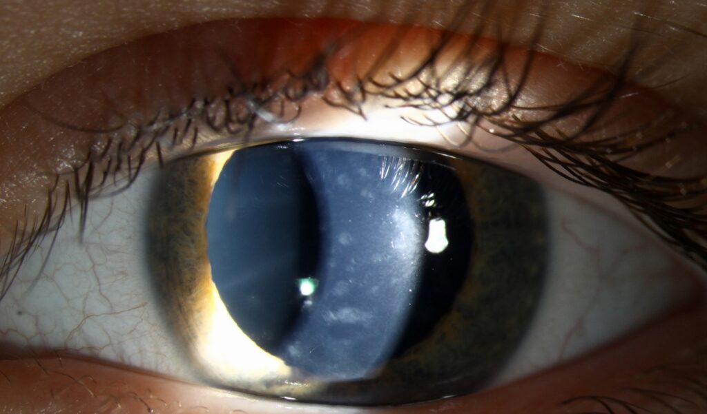
Cornea farinata is a benign, age-related pattern of tiny gray dots in the deep corneal stroma.
The name comes from the Latin word for flour, because the opacities resemble a fine dusting of flour. It is usually bilateral and found by chance during a routine slit-lamp examination, and vision is unaffected, so the main task is correct recognition and calm reassurance.
What Cornea Farinata Looks Like
The dots are minute, uniform and gray-white, scattered across the posterior stroma, and they sit just in front of Descemet membrane and are often densest centrally. Each dot lies near the limit of slit-lamp resolution, and as a result, a quick direct beam often misses them entirely.
Retroillumination for Cornea Farinata
Retroillumination from the iris or the red reflex shows the dots best, and they then appear as countless fine specks, like dust on a window pane. Next, a narrow tangential slit localizes them to the pre-Descemet layer, and higher magnification confirms that the epithelium and anterior stroma remain clear.
All-fit Slit-Lamp Adapter
Record and share exactly what you see at the slit lamp. One adapter fits any slit lamp or surgical microscope — and any smartphone.
From Choroida — the team behind this siteWhy the Dots Form
The exact origin of the opacities is uncertain, and histologic reports of related pre-Descemet changes describe vacuolated posterior keratocytes with lipofuscin-like material. Therefore, most authors regard the finding as a degenerative aging change.
Who Gets Cornea Farinata
It appears mainly in older adults, typically after middle age, and both sexes are affected, with both eyes usually showing a similar pattern. Occasional families show similar deep dots, which suggests some overlap with dystrophy. However, most patients have no family history and no linked systemic disease.
Symptoms and Vision
The dots themselves cause no symptoms, and visual acuity, contrast and corneal thickness stay normal. Therefore, any blur needs another explanation, such as cataract, dry eye or macular disease.
Where It Sits in Classification
The IC3D classification discusses cornea farinata alongside pre-Descemet corneal dystrophy, and some authors view it as a mild age-related variant, while others call it a separate degeneration. In practice, the label changes little, because both are benign (see pre-Descemet corneal dystrophy).
Cornea Farinata Versus Pre-Descemet Dystrophy
Pre-Descemet dystrophy often shows larger, polymorphic opacities with comma, dendritic or annular shapes, and it may also occur with X-linked ichthyosis, so a skin history is useful. In contrast, farinata dots are tiny, uniform and dust-like.
Telling Cornea Farinata From Guttae
Guttae are excrescences of Descemet membrane produced by stressed endothelium, and on specular reflection, they appear as dark spots that interrupt the endothelial mosaic. Later, they coalesce into a beaten-metal pattern and may cause stromal edema. Farinata dots, however, lie in the stroma and leave the endothelial reflex intact, so specular microscopy settles doubtful cases (see Fuchs endothelial dystrophy).
Other Deep Corneal Look-Alikes
- Polymorphic amyloid degeneration: refractile polygonal flecks in the deep stroma of older adults
- Posterior crocodile shagreen: gray polygonal mosaic separated by clear lines
- Fleck dystrophy: discrete flecks at all stromal levels, sometimes asymmetric
- Keratic precipitates: endothelial deposits with cells or flare in the anterior chamber
- Pigment dusting: brown granules on the endothelium, often in a vertical spindle
Examination Steps
First, confirm that both eyes show similar, symmetric dots, and then check the depth with a thin slit and high magnification. Also look for epithelial disease, stromal haze, edema or inflammation, and finally, review the specular reflex for guttae.
Imaging and Tests
Most patients need no investigation at all, but when the depth is unclear, specular microscopy usually shows normal endothelial morphology. Pachymetry is also normal because the dots do not affect corneal hydration, while confocal microscopy has been reported to show hyperreflective particles in posterior keratocytes.
Does Cornea Farinata Need Treatment?
No treatment is required, and drops, ointments and dietary changes have no role. The dots appear stable over long periods, so routine eye examinations are sufficient.
Cornea Farinata and Eye Surgery
The finding does not contraindicate cataract or refractive surgery. However, document it clearly before any procedure, because otherwise a later examiner may attribute the deep dots to surgical trauma or toxicity.
Counseling Patients With Cornea Farinata
Explain that the dots are a common aging change in the back layer of the cornea. Then reassure the patient that they do not affect vision or progress to blindness, and also explain that they are not infectious, not inflammatory and need no drops.
When to Look Again
Re-examine the cornea if new blur, glare or pain develops. Similarly, asymmetric dots, enlarging opacities or morning haze suggest another diagnosis, so in that case, reassess for endothelial disease, inflammation or dystrophy.
Pitfalls to Avoid
- Do not miss the dots by using only a broad direct beam
- Never label stromal dots as guttae without checking specular reflection
- Do not order extensive tests for a classic bilateral, symmetric pattern
- Avoid blaming farinata for reduced vision
- Always record the finding before intraocular surgery


Document what you see
Two smartphone imaging tools built for everyday clinic use — one for the slit lamp, one for the fundus.
From Choroida — the team behind this siteReferences
- Weiss JS, Møller HU, Aldave AJ, et al. IC3D classification of corneal dystrophies, edition 2. Cornea. 2015;34:117-159.
- Curran RE, Kenyon KR, Green WR. Pre-Descemet’s membrane corneal dystrophy. Am J Ophthalmol. 1974;77:711-716.
- Salmon JF. Kanski’s Clinical Ophthalmology: A Systematic Approach. 9th ed. Elsevier; 2020.
- Mannis MJ, Holland EJ. Cornea. 4th ed. Elsevier; 2017.
- American Academy of Ophthalmology. Basic and Clinical Science Course, Section 8: External Disease and Cornea. AAO; 2023.
Test yourself
A few questions straight from this article.
-
Where do the dots of cornea farinata lie?
The dots are minute, uniform gray-white specks in the posterior stroma just in front of Descemet membrane, often densest centrally. -
Which slit lamp technique shows the dots of cornea farinata best?
Each dot lies near the limit of slit lamp resolution, so a quick direct beam often misses them; retroillumination shows them as countless fine specks. -
What do histologic reports of related pre-Descemet changes describe as the likely basis of cornea farinata?
The exact origin is uncertain, but reports describe vacuolated posterior keratocytes with lipofuscin-like material, so most authors regard it as a degenerative aging change. -
A patient with cornea farinata reports blurred vision. How should this be interpreted?
The dots cause no symptoms and leave acuity, contrast and thickness normal, so any blur needs another explanation such as cataract, dry eye or macular disease. -
Which systemic association points to pre-Descemet dystrophy rather than cornea farinata?
Pre-Descemet dystrophy shows larger polymorphic opacities and may occur with X-linked ichthyosis, so a skin history helps; farinata dots are tiny, uniform and dust-like. -
How are the dots of cornea farinata distinguished from corneal guttae?
Guttae appear as dark spots interrupting the endothelial mosaic; farinata dots lie in the stroma and leave the endothelial reflex intact, so specular microscopy settles doubtful cases. -
What treatment does cornea farinata require?
No treatment is required and drops, ointments and dietary changes have no role; the dots appear stable over long periods. -
Why should cornea farinata be documented before cataract or refractive surgery?
Farinata does not contraindicate surgery, but if undocumented a later examiner may attribute the deep dots to surgical trauma or toxicity. -
In a patient thought to have cornea farinata, which finding suggests another diagnosis?
Asymmetric dots, enlarging opacities or morning haze suggest another diagnosis and prompt reassessment for endothelial disease, inflammation or dystrophy. -
Why is pachymetry normal in cornea farinata?
Pachymetry is normal because the dots do not affect corneal hydration, and specular microscopy usually shows normal endothelial morphology.

Writes and reviews the clinical articles on Educate. Every post is checked against peer-reviewed sources before it goes live.