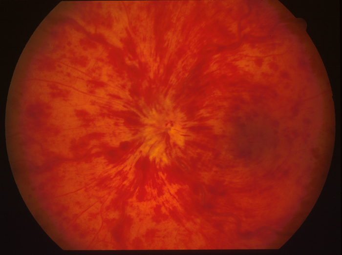
Blood and thunder fundus is the classic description of severe central retinal vein occlusion (CRVO).
The examiner sees hemorrhages in all four quadrants, dilated tortuous veins, and often a swollen disc. However, the name describes an appearance and does not establish perfusion status, so each case needs a structured search for ischemia.
What Blood and Thunder Looks Like
Flame-shaped hemorrhages radiate from the disc along the nerve fiber layer, and deeper dot and blot hemorrhages also scatter across the posterior pole and periphery. The veins look dark and engorged, while the disc margin often blurs, and cotton wool spots and macular edema frequently complete the picture.
Why the Hemorrhages Appear
Thrombosis at or near the lamina cribrosa obstructs venous outflow from the whole retina. As a result, venous and capillary pressure rise, vessels leak blood and fluid, and sluggish circulation can lead to capillary closure.
Fundus Explorer Pro
Photograph the retinal findings described here with the phone already in your pocket — 22 D optics and built-in illumination in one handheld unit.
From Choroida — the team behind this siteWhat Patients Report
Many patients notice painless vision loss in one eye, sometimes on waking, and some describe sudden blur while others report a decline over days. Pain and redness suggest a later complication, particularly neovascular glaucoma, while earlier transient blur may precede a completed occlusion, so ask about it.
Does Blood and Thunder Mean Ischemia?
The appearance raises suspicion of ischemia but cannot confirm it, because extensive hemorrhage can also occur in nonischemic CRVO, particularly early. Therefore, judge perfusion from several tests together (see ischemic CRVO).
Bedside Signs of Ischemia
First, check visual acuity, because very poor vision increases concern. Next, test the pupils, since a relative afferent defect supports ischemic disease. Finally, note numerous cotton wool spots, although their absence does not exclude ischemia.
Imaging a Blood and Thunder Fundus
Widefield fluorescein angiography maps capillary nonperfusion and leakage, but dense hemorrhage blocks fluorescence, so an early angiogram may be indeterminate. The Central Vein Occlusion Study (CVOS) defined nonperfusion as at least 10 disc areas of capillary closure, while OCT measures macular edema and OCT angiography shows macular capillary loss without dye. Therefore, repeat angiography when the hemorrhage clears.
Electroretinography in CRVO
Electroretinography assesses retinal function when hemorrhage obscures the view, and a reduced b-wave amplitude and low b/a ratio support ischemia. Because no single test settles the question, interpret results alongside clinical findings.
What to Check Next
First, measure intraocular pressure and examine the undilated iris at high magnification. Next, perform gonioscopy before dilation, because angle vessels can appear before iris vessels, and then examine the fellow eye and check blood pressure.
Neovascular Risk After Blood and Thunder
CVOS linked poor initial acuity and nonperfusion with a higher risk of iris neovascularization. Perfusion status can also change, and some perfused eyes later became nonperfused, so one reassuring angiogram does not end monthly surveillance in high-risk eyes.
Blood and Thunder Differential Diagnosis
Ocular ischemic syndrome causes midperipheral hemorrhages with dilated, less tortuous veins. Bilateral disease should prompt evaluation for hyperviscosity, severe hypertension or leukemic retinopathy, and diabetic and radiation retinopathy also enter the differential when the history fits.
Systemic Workup
Common associations include hypertension, diabetes and glaucoma, so coordinate vascular risk assessment with the primary physician. However, routine thrombophilia panels add little for older patients with typical risk factors.
Treating Macular Edema in Blood and Thunder
Anti-VEGF injections reduce vascular permeability and treat macular edema. For example, ranibizumab improved six-month vision more than sham in CRUISE, and SCORE2 also found bevacizumab noninferior to aflibercept at six months. However, injections cannot restore a macula with established capillary loss and carry a small endophthalmitis risk, so tell patients that treatment often requires repeated visits.
Panretinal Photocoagulation
Panretinal photocoagulation (PRP) ablates ischemic retina and reduces angiogenic drive, but prophylactic PRP did not reliably prevent neovascularization in CVOS. Therefore, apply PRP promptly once iris or angle neovascularization appears, and because PRP can reduce peripheral and night vision, discuss these effects first. Anti-VEGF may temporarily suppress new vessels, so surveillance should continue after injections stop.
Prognosis and Follow-Up
Eyes with good initial vision tended to keep it in CVOS, whereas eyes starting at 20/200 or worse seldom recovered substantially. Advise urgent review for pain, redness or a further drop in vision, and distinguish this picture from arterial disease, which demands a different pathway (see CRAO vs CRVO).
Pitfalls to Avoid
- Do not equate hemorrhage volume with confirmed ischemia
- Avoid relying on an early angiogram blocked by hemorrhage
- Do not dilate before examining the iris and angle
- Never assume bilateral hemorrhages represent simple CRVO
- Do not stop neovascular surveillance after anti-VEGF therapy ends


Document what you see
Two smartphone imaging tools built for everyday clinic use — one for the slit lamp, one for the fundus.
From Choroida — the team behind this siteReferences
- The Central Vein Occlusion Study Group. Natural history and clinical management of central retinal vein occlusion. Arch Ophthalmol. 1997;115:486-491.
- The Central Vein Occlusion Study Group. A randomized clinical trial of early panretinal photocoagulation for ischemic central vein occlusion: the CVOS Group N report. Ophthalmology. 1995;102:1434-1444.
- Brown DM, Campochiaro PA, Singh RP, et al. Ranibizumab for macular edema following central retinal vein occlusion: six-month primary end point results of a phase III study. Ophthalmology. 2010;117:1124-1133.
- Scott IU, VanVeldhuisen PC, Ip MS, et al. Effect of bevacizumab vs aflibercept on visual acuity among patients with macular edema due to central retinal vein occlusion: the SCORE2 randomized clinical trial. JAMA. 2017;317:2072-2087.
- Flaxel CJ, Adelman RA, Bailey ST, et al. Retinal Vein Occlusions Preferred Practice Pattern. Ophthalmology. 2020;127:P288-P320.
Test yourself
A few questions straight from this article.
-
Which fundus picture is described as a 'blood and thunder' fundus in central retinal vein occlusion?
Severe CRVO shows hemorrhages in all four quadrants, dark engorged tortuous veins and often a swollen disc, giving the blood and thunder appearance. -
Where does the thrombosis that produces a blood and thunder fundus in CRVO typically occur?
Thrombosis at or near the lamina cribrosa obstructs venous outflow from the whole retina, raising venous and capillary pressure so vessels leak blood and fluid. -
Does a blood and thunder fundus in CRVO confirm ischemic disease?
The appearance raises suspicion of ischemia but cannot confirm it, because extensive hemorrhage can occur in nonischemic CRVO, especially early, so perfusion is judged from several tests together. -
Which bedside pupil finding supports ischemic central retinal vein occlusion?
Alongside very poor acuity and numerous cotton wool spots, a relative afferent pupillary defect supports ischemic CRVO. -
How did the Central Vein Occlusion Study (CVOS) define nonperfused CRVO on fluorescein angiography?
CVOS defined nonperfusion as at least 10 disc areas of capillary closure; dense hemorrhage can block an early angiogram, so it is repeated as the blood clears. -
Which electroretinography result supports ischemia in CRVO when hemorrhage obscures the fundus view?
Electroretinography assesses retinal function when the view is poor; a reduced b-wave amplitude and low b/a ratio support ischemia, interpreted alongside clinical findings. -
In CRVO, why should gonioscopy be performed before pupil dilation?
Intraocular pressure and the undilated iris are examined first, and gonioscopy precedes dilation because angle neovascularization can appear before iris vessels. -
Which fundus pattern points to ocular ischemic syndrome rather than a blood and thunder CRVO?
Ocular ischemic syndrome causes midperipheral hemorrhages with dilated but less tortuous veins, unlike the disc-centred four-quadrant picture of CRVO. -
In central retinal vein occlusion, when should panretinal photocoagulation be applied?
Prophylactic PRP did not reliably prevent neovascularization in CVOS, so PRP is applied promptly once iris or angle new vessels appear. -
In CVOS, which presenting visual acuity in CRVO predicted little substantial recovery?
Eyes with good initial vision tended to keep it, whereas eyes starting at 20/200 or worse seldom recovered substantially; poor initial acuity also raised iris neovascularization risk.

Writes and reviews the clinical articles on Educate. Every post is checked against peer-reviewed sources before it goes live.