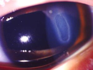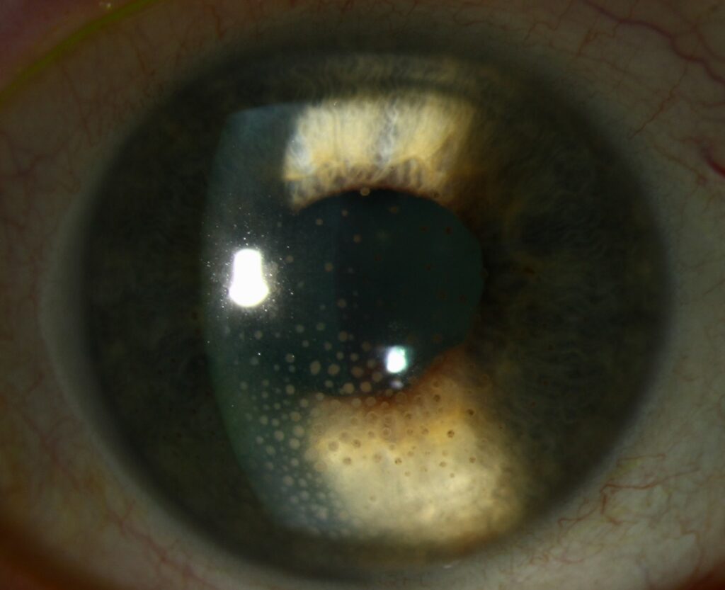
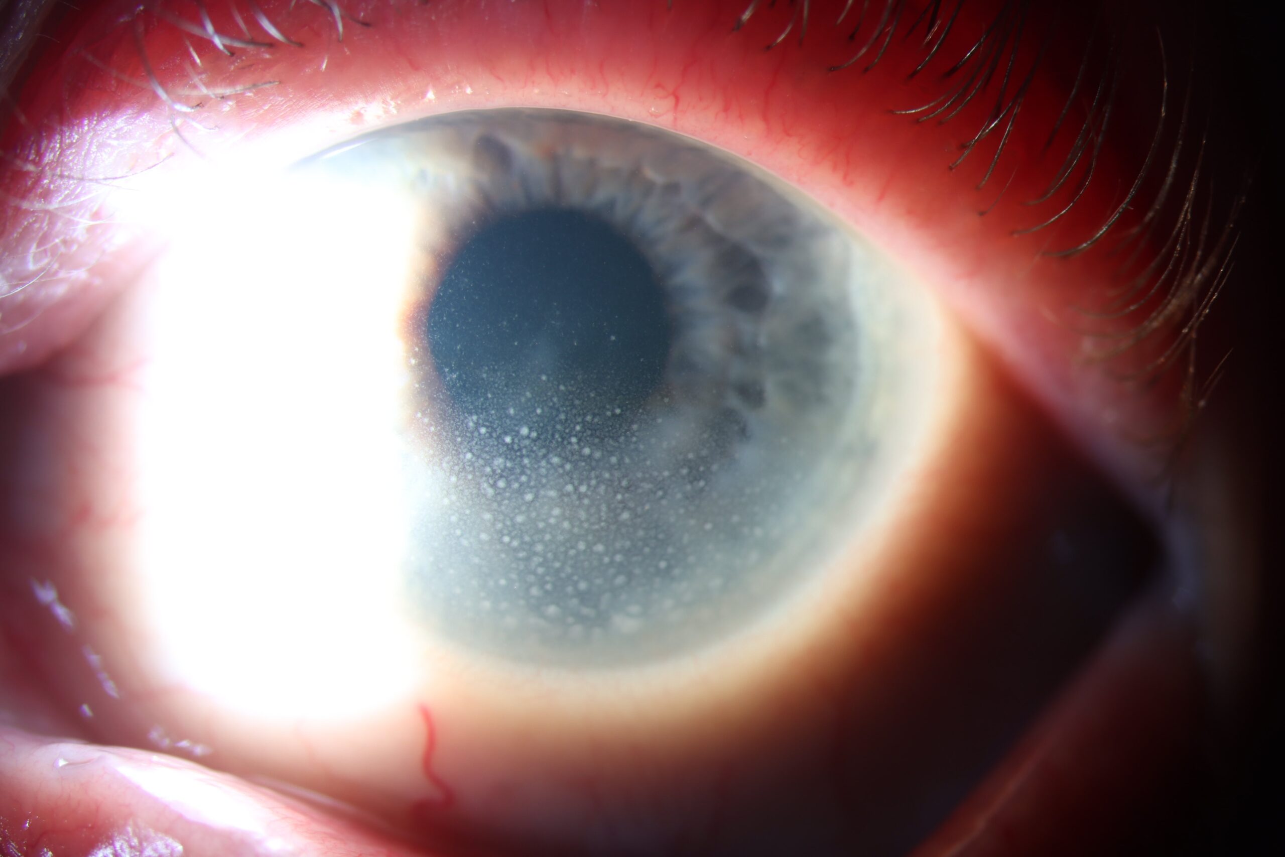
Mutton fat KP are large, greasy keratic precipitates on the corneal endothelium, typically seen in granulomatous anterior uveitis.
They contain macrophages and epithelioid cells, so they look bulkier than fine nongranulomatous deposits. Therefore, this sign should prompt a targeted search for infection and systemic disease.
What Mutton Fat KP Look Like
These deposits appear yellow-white, rounded and waxy, with soft, raised margins, and their size often varies within the same eye. Older deposits become pigmented, flatter and glassy as inflammation settles, although patients usually report blur, redness or photophobia from the uveitis itself.
Distribution of Mutton Fat KP
Inflammatory cells circulate in the aqueous, then adhere to the inflamed endothelium. Convection currents and gravity favor the inferior cornea, a zone called Arlt’s triangle, so a base-down triangular pattern is common. Diffuse deposits, by contrast, should prompt consideration of Fuchs uveitis syndrome or herpetic disease.
All-fit Slit-Lamp Adapter
Record and share exactly what you see at the slit lamp. One adapter fits any slit lamp or surgical microscope — and any smartphone.
From Choroida — the team behind this siteSlit Lamp Technique
Begin with a broad beam, then narrow it to assess the endothelium (see slit lamp exam), while retroillumination also helps separate true deposits from pigment dusting and guttae. Next, record their size, color and distribution, and finally photograph them when possible, because serial images make comparison easier.
Supporting Signs
Koeppe nodules at the pupil margin and Busacca nodules on the iris strengthen the impression, and broad posterior synechiae suggest long-standing inflammation. A dilated examination then checks for vitritis, snowballs, periphlebitis and choroidal lesions.
Main Causes of Mutton Fat KP
- Sarcoidosis
- Tuberculosis
- Syphilis
- Vogt-Koyanagi-Harada disease
- Sympathetic ophthalmia
- Lens-induced uveitis
- Herpetic uveitis
- Toxoplasmosis
- Chronic postoperative endophthalmitis
Sarcoidosis, Tuberculosis and Syphilis
Sarcoid uveitis is often bilateral and may produce iris nodules, snowballs and periphlebitis, while the revised international criteria also include multifocal choroidal lesions and tent-shaped peripheral anterior synechiae. However, these signs do not establish the diagnosis alone.
Tuberculosis may cause broad synechiae, retinal vasculitis or serpiginous-like choroiditis. Therefore, test for syphilis and tuberculosis in almost every patient, because treatment differs.
Vogt-Koyanagi-Harada and Sympathetic Ophthalmia
Both conditions cause bilateral panuveitis, so the posterior segment often carries the diagnosis, and Vogt-Koyanagi-Harada disease may include serous detachments, headache and tinnitus. In contrast, sympathetic ophthalmia follows penetrating trauma or surgery in the fellow eye, so always ask about remote injuries.
Mutton Fat KP After Surgery or Trauma
Low-grade inflammation after cataract surgery raises concern for chronic endophthalmitis, including Cutibacterium acnes. Look carefully for a white capsular plaque, because steroids may only partly suppress this infection. Retained lens material can also drive granulomatous inflammation, and when infection remains plausible, vitreous or aqueous sampling may guide treatment.
Conditions That Can Mislead
Herpetic uveitis often pairs deposits with sectoral iris atrophy, corneal scars or raised pressure. Similarly, high pressure with few small deposits suggests a different diagnosis (see Posner-Schlossman syndrome). Fuchs uveitis syndrome, meanwhile, produces fine, stellate deposits across the endothelium.
Tests Prompted by Mutton Fat KP
Testing should follow the clinical picture, although a few baseline tests apply widely.
- Treponemal syphilis serology
- Interferon-gamma release assay or tuberculin skin test
- Chest imaging, with CT when suspicion persists
- Aqueous PCR for suspected herpetic disease
- OCT and fluorescein angiography for posterior involvement
A positive tuberculosis test does not prove ocular infection, and likewise a normal angiotensin-converting enzyme level does not exclude sarcoidosis.
Treating Mutton Fat KP
Treatment targets the cause, since the deposits only mark inflammation, and topical corticosteroids reduce anterior inflammation, while cycloplegics ease ciliary spasm and prevent synechiae. However, steroids cannot treat infection, and they may worsen it without antimicrobial cover.
They also raise pressure and promote cataract, so check pressure at each visit, and systemic immunosuppression may follow for bilateral, sight-threatening noninfectious disease.
Follow-Up and Disease Activity
Deposits can persist after active inflammation has resolved, so base decisions on anterior chamber cells and the overall examination. New white deposits suggest activity, while pigmented, flattened ones suggest older disease, and also check the macula for cystoid macular edema.
Counseling Patients About Mutton Fat KP
Explain that these deposits are clusters of inflammatory cells on the inner corneal surface, then clarify that they point toward a type of inflammation, without naming one specific disease. Patients should also know why blood tests and chest imaging may be requested, and finally, they should never stop steroid drops abruptly.
Pitfalls to Avoid
- Do not assume noninfectious disease before excluding syphilis and tuberculosis
- Do not treat chronic postoperative inflammation with steroids alone
- Never skip the dilated fundus examination
- Do not equate old deposits with active uveitis


Document what you see
Two smartphone imaging tools built for everyday clinic use — one for the slit lamp, one for the fundus.
From Choroida — the team behind this siteReferences
- Jabs DA, Nussenblatt RB, Rosenbaum JT, et al. Standardization of uveitis nomenclature for reporting clinical data. Am J Ophthalmol. 2005;140:509-516.
- Mochizuki M, Smith JR, Takase H, et al. Revised criteria of International Workshop on Ocular Sarcoidosis (IWOS) for the diagnosis of ocular sarcoidosis. Br J Ophthalmol. 2019;103:1418-1422.
- Gupta V, Gupta A, Rao NA. Intraocular tuberculosis, an update. Surv Ophthalmol. 2007;52:561-587.
- Read RW, Holland GN, Rao NA, et al. Revised diagnostic criteria for Vogt-Koyanagi-Harada disease. Am J Ophthalmol. 2001;131:647-652.
Test yourself
A few questions straight from this article.
-
Mutton fat keratic precipitates are typically seen in which type of uveitis?
Mutton fat KP are large, greasy keratic precipitates on the corneal endothelium, typically seen in granulomatous anterior uveitis. -
Which cells make mutton fat keratic precipitates bulkier than fine nongranulomatous deposits?
Mutton fat KP contain macrophages and epithelioid cells, so they look bulkier than fine nongranulomatous deposits. -
Where on the cornea do mutton fat keratic precipitates typically collect?
Convection currents and gravity favor the inferior cornea, a zone called Arlt's triangle, so a base-down triangular pattern is common. -
How do the keratic precipitates of Fuchs uveitis syndrome differ from mutton fat KP?
Fuchs uveitis syndrome produces fine, stellate deposits across the endothelium, so diffuse deposits should prompt consideration of it rather than a granulomatous cause. -
Which nodules at the pupil margin support a granulomatous cause alongside mutton fat KP?
Koeppe nodules at the pupil margin and Busacca nodules on the iris strengthen the impression, and broad posterior synechiae suggest long-standing inflammation. -
Low-grade inflammation with mutton fat KP after cataract surgery and a white capsular plaque suggests which organism?
Low-grade postoperative inflammation raises concern for chronic endophthalmitis, including Cutibacterium acnes; look for a white capsular plaque, because steroids may only partly suppress it. -
Bilateral panuveitis with mutton fat KP after penetrating trauma to one eye suggests which diagnosis?
Both VKH and sympathetic ophthalmia cause bilateral panuveitis, but sympathetic ophthalmia follows penetrating trauma or surgery in the fellow eye, so always ask about remote injuries. -
Which two infections should be tested for in almost every patient with mutton fat KP?
Syphilis and tuberculosis should be tested in almost every patient because treatment differs, and noninfectious disease should not be assumed before excluding them. -
In a patient with mutton fat KP, what does a normal angiotensin-converting enzyme level mean?
A normal angiotensin-converting enzyme level does not exclude sarcoidosis, just as a positive tuberculosis test does not prove ocular infection. -
At follow-up, which mutton fat KP appearance suggests active inflammation?
Deposits can persist after inflammation resolves, so decisions rest on anterior chamber cells; new white deposits suggest activity, while pigmented, flattened ones suggest older disease.

Writes and reviews the clinical articles on Educate. Every post is checked against peer-reviewed sources before it goes live.