Ulcer vs abrasion is a common triage question because both lesions cause pain, redness and fluorescein uptake.
An abrasion is a mechanical epithelial defect, while an ulcer adds stromal inflammation, usually from infection.
Therefore, the examiner must look beneath the stain.
What Separates Ulcer vs Abrasion
An abrasion removes epithelium and leaves the underlying stroma largely clear.
In contrast, an ulcer combines an epithelial defect with a stromal infiltrate, sometimes with tissue loss.
Microbes, immune disease or neurotrophic damage can all produce this picture.
As a result, the two lesions carry very different risks to vision.
History in Ulcer vs Abrasion
Abrasion patients usually recall a precise event, such as a fingernail, paper edge or branch.
Pain then starts immediately and typically eases as the epithelium closes.
Ulcer patients often report contact lens wear, and their pain, discharge and blur worsen over time.
Schein and colleagues found a higher risk of ulcerative keratitis with extended wear than with daily wear.
All-fit Slit-Lamp Adapter
Record and share exactly what you see at the slit lamp. One adapter fits any slit lamp or surgical microscope — and any smartphone.
From Choroida — the team behind this siteThe Infiltrate as the Deciding Sign
A stromal infiltrate appears as a white or gray opacity with hazy borders.
It forms because inflammatory cells migrate into the stroma.
A pure abrasion should lack this density, although mild adjacent edema can occur.
However, small peripheral lesions may be sterile (see sterile versus infectious infiltrates).
Staining in Ulcer vs Abrasion
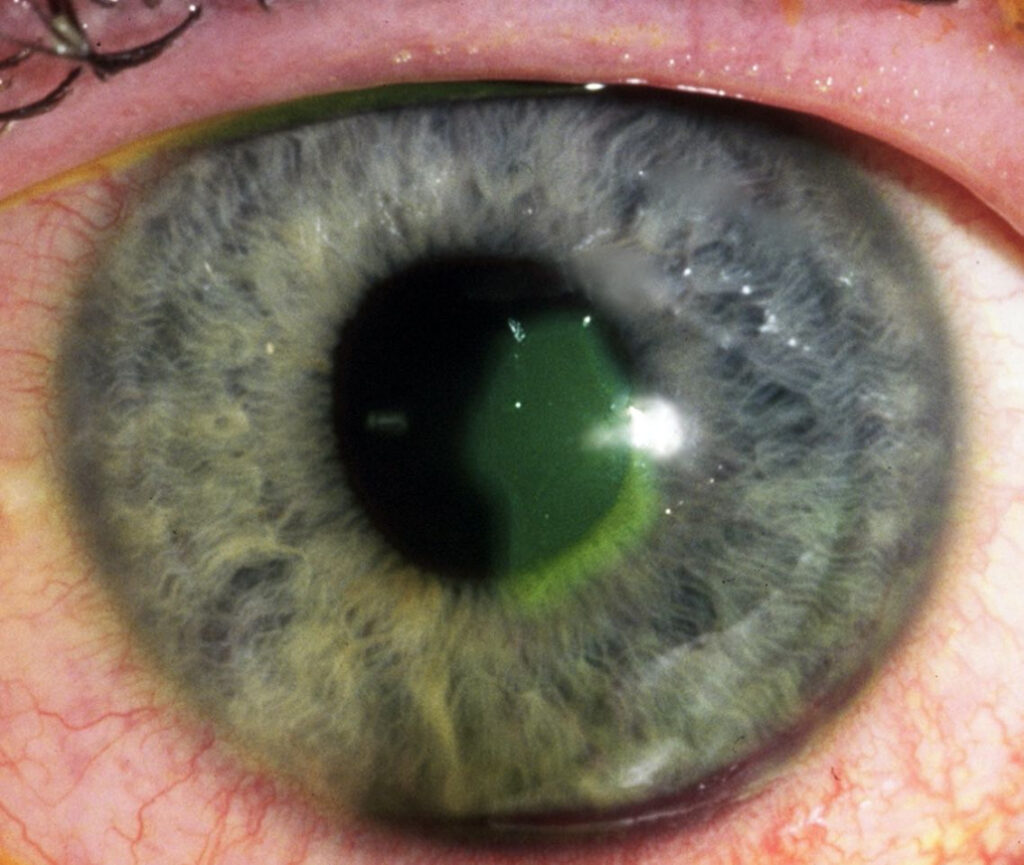
Fluorescein marks missing epithelium in both lesions, so staining alone cannot separate them.
An abrasion typically shows sharp margins over clear stroma.
Vertical linear streaks also suggest a foreign body under the upper lid.
An ulcer usually stains over a white bed, often with irregular edges.
In addition, a branching pattern with terminal bulbs points to herpes simplex keratitis.
Urgency in Ulcer vs Abrasion
Clinicians can usually manage a small traumatic abrasion without infiltrate on an outpatient basis.
Any suspected ulcer, however, needs same day slit lamp assessment.
Hypopyon strongly favors an ulcer.
Contact lens wearers need a low threshold for urgent review because Pseudomonas can progress rapidly.
Similarly, vegetable matter trauma raises concern for fungal infection.
Applying the 1, 2, 3 Rule
Vital and colleagues proposed a simple rule for grading ulcer severity.
It flags 1+ anterior chamber cells, an infiltrate of 2 mm, or a location within 3 mm of the visual axis.
Any positive criterion supports culture and closer care.
However, smaller lesions can still behave aggressively.
When to Culture
AAO guidance favors smears and cultures for large, central or deep infiltrates.
Chronic, unresponsive or atypical lesions also merit sampling.
Whenever feasible, take scrapings before antibiotics, without causing undue delay.
A clean abrasion needs no culture unless an infiltrate develops.
Treating an Abrasion
Typical care includes lubrication, oral analgesia and a topical antibiotic, although evidence for prophylaxis is limited.
A Cochrane review found that patching did not speed healing or reduce pain (see patching and drops).
Contact lens wearers should stop lens wear and receive antipseudomonal cover.
Even so, drops cannot prevent every later recurrent erosion.
Treating an Ulcer
Frequent broad spectrum topical antibiotics form initial therapy, using a fluoroquinolone or fortified combination.
Clinicians then adjust treatment according to culture results and clinical response.
Antibiotics can eradicate organisms, but they cannot reverse established scarring.
Therefore, warn patients with central lesions about possible permanent blur.
Steroids and Infection Risk
Never start a steroid for an undifferentiated painful red eye, because it can worsen herpetic epithelial and fungal disease.
SCUT tested adjunctive prednisolone after 48 hours of moxifloxacin in culture positive bacterial ulcers.
It found no overall benefit in 3 month visual acuity (see the SCUT trial).
Follow-Up in Ulcer vs Abrasion
Recheck large, central or lens related abrasions within about 24 hours.
If a defect stops shrinking or a white opacity appears, reclassify the lesion as an ulcer.
Meanwhile, review significant ulcers daily until they clearly improve.
Counseling in Ulcer vs Abrasion
Tell abrasion patients that discomfort should improve each day.
They should return immediately for rising pain, falling vision, discharge or a white spot.
Also warn them about recurrent erosion, which often causes sudden pain on waking.
Ulcer patients need strict dosing, including night doses when prescribed, and no lens wear.
Pitfalls to Avoid
- Do not call every fluorescein defect an abrasion before checking the stroma
- Never prescribe topical anesthetic drops for home use
- Avoid patching lens wearers or any eye with suspected infection
- Do not dismiss a quiet white lesion in a cornea with reduced sensation


Document what you see
Two smartphone imaging tools built for everyday clinic use — one for the slit lamp, one for the fundus.
From Choroida — the team behind this siteReferences
- Lin A, Rhee MK, Akpek EK, et al. Bacterial Keratitis Preferred Practice Pattern. Ophthalmology. 2019;126:P1-P55.
- Vital MC, Belloso M, Prager TC, Lanier JD. Classifying the severity of corneal ulcers by using the “1, 2, 3” rule. Cornea. 2007;26:16-20.
- Srinivasan M, Mascarenhas J, Rajaraman R, et al. Corticosteroids for bacterial keratitis: the Steroids for Corneal Ulcers Trial (SCUT). Arch Ophthalmol. 2012;130:143-150.
- Turner A, Rabiu M. Patching for corneal abrasion. Cochrane Database Syst Rev. 2006;(2):CD004764.
- Wipperman JL, Dorsch JN. Evaluation and management of corneal abrasions. Am Fam Physician. 2013;87:114-120.
- Schein OD, Glynn RJ, Poggio EC, et al. The relative risk of ulcerative keratitis among users of daily-wear and extended-wear soft contact lenses. N Engl J Med. 1989;321:773-778.
Test yourself
A few questions straight from this article.
-
Which finding converts a fluorescein-staining corneal epithelial defect from an abrasion into an ulcer?
An abrasion is a mechanical epithelial defect over largely clear stroma, while an ulcer adds a white or gray stromal infiltrate from migrating inflammatory cells. -
Why can fluorescein staining alone not separate a corneal ulcer from an abrasion?
Both lesions lack epithelium and stain, so the examiner must look beneath the stain for a stromal infiltrate. -
Vertical linear fluorescein streaks on the cornea suggest what cause?
An abrasion typically stains with sharp margins over clear stroma, and vertical linear streaks point to a foreign body under the upper lid. -
A branching corneal staining pattern with terminal bulbs points to which diagnosis?
A branching pattern with terminal bulbs points to herpes simplex keratitis, which steroids can worsen. -
Which anterior segment sign strongly favors a corneal ulcer over an abrasion?
Hypopyon strongly favors an ulcer, and any suspected ulcer needs same-day slit lamp assessment. -
How should a corneal abrasion in a contact lens wearer be managed?
Pseudomonas can progress rapidly in lens wearers, so they stop lens wear, receive antipseudomonal cover, and are not patched. -
In the '1, 2, 3' rule for corneal ulcer severity, what does the number 3 refer to?
The rule flags anterior chamber cells, infiltrate size, and a location within 3 mm of the visual axis; any positive criterion supports culture and closer care. -
When should corneal scrapings for smear and culture be taken in suspected microbial keratitis?
Smears and cultures are favored for large, central or deep infiltrates, taken before antibiotics whenever feasible without delaying treatment. -
What did a Cochrane review conclude about patching a corneal abrasion?
Patching did not speed healing or reduce pain; abrasion care is lubrication, oral analgesia and a topical antibiotic. -
In the Steroids for Corneal Ulcers Trial (SCUT), what did adjunctive prednisolone add in culture-positive bacterial ulcers?
SCUT added prednisolone after 48 hours of moxifloxacin and found no overall benefit in 3-month acuity; steroids are never started for an undifferentiated red eye.

Writes and reviews the clinical articles on Educate. Every post is checked against peer-reviewed sources before it goes live.