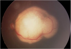
Retinal whitening is an opaque, pale change in retinal tissue that usually reflects ischemia, mechanical injury or necrosis.
The healthy retina is nearly transparent, so the choroidal color normally shows through.
Therefore, any loss of transparency stands out on fundus examination.
Distribution, borders and vessel changes then narrow the differential quickly.
Why Retinal Whitening Appears
Ischemia causes intracellular swelling of the inner retina, which scatters light and hides the choroid.
In contrast, blunt trauma mainly disrupts the photoreceptor outer segments.
Infection produces whitening through retinal necrosis, often with adjacent vascular disease.
As a result, similar pallor can arise from very different tissue events.
Reading Retinal Whitening by Pattern
Start with four questions at the slit lamp:
- Does the lesion fill a vascular territory?
- Is its edge sharp, feathery or granular?
- Do hemorrhages or sheathed vessels accompany it?
- Is the vitreous clear or inflamed?
In addition, ask about onset, pain, recent trauma and immune status.
Fundus Explorer Pro
Photograph the retinal findings described here with the phone already in your pocket — 22 D optics and built-in illumination in one handheld unit.
From Choroida — the team behind this siteDiffuse Retinal Whitening in CRAO
Central retinal artery occlusion causes sudden, painless, severe vision loss, often with a relative afferent pupillary defect.
The posterior pole turns white, while the fovea shows a cherry red spot because its thin retina lets the choroid show through.
However, whitening can take hours to develop, so an early normal fundus does not exclude occlusion.
Treat this presentation as a stroke equivalent and arrange urgent vascular evaluation.
Also, exclude giant cell arteritis in older patients.
Sectoral Whitening and Branch Occlusion
A sharply bordered pale zone within one arterial territory suggests branch retinal artery occlusion.
Next, inspect the affected artery for an embolus, especially at a bifurcation.
Patients usually report a sudden field defect that matches the pale sector.
Cilioretinal artery occlusion looks similar, although its whitening stays near the papillomacular region.
Band-Like Whitening in PAMM
Paracentral acute middle maculopathy appears as subtle gray-white parafoveal bands.
OCT shows a hyperreflective band at the inner nuclear layer, which reflects capillary ischemia.
Patients often describe a sudden paracentral scotoma despite good acuity.
Therefore, search for vascular risk factors and an associated retinal vascular occlusion.
Multifocal Patches and Purtscher Retinopathy
Cotton wool spots are small, feathery, superficial lesions caused by arrested axoplasmic flow.
They commonly accompany hypertension and diabetes, and they also occur in HIV retinopathy.
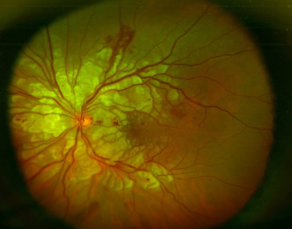
Purtscher retinopathy produces larger polygonal patches between arterioles, usually around the disc.
For example, it can follow head or chest trauma, or acute pancreatitis.
No proven treatment exists, so management targets the underlying condition.
Retinal Whitening After Blunt Trauma
Commotio retinae causes gray-white outer retinal opacification within hours of blunt injury.
The lesion ignores arterial territories, and the vessels look normal.
OCT shows disturbed outer retinal reflectivity, while the inner retina stays largely intact.
Most cases clear over days to weeks.
However, macular involvement can leave permanent photoreceptor loss, so counsel patients cautiously.
High-Velocity Injury and Sclopetaria
A projectile passing near the globe can cause extensive retinal and choroidal disruption without scleral rupture.
Retinitis sclopetaria combines whitening, hemorrhage and later claw-like scarring.
Consequently, always exclude an open globe and intraocular foreign body first.
Observation is usual, because the scar often limits the risk of detachment.
Peripheral Retinal Whitening in Acute Necrosis
Acute retinal necrosis starts with peripheral patches that merge and advance circumferentially.
Occlusive arteritis and prominent vitritis complete the classic picture.
Because progression can be rapid, send intraocular fluid for herpesvirus PCR and start antiviral therapy promptly.
Antivirals can limit spread and may protect the fellow eye, but they cannot restore necrotic retina.
Warn patients about later retinal detachment.
Granular Retinal Whitening in CMV
Cytomegalovirus retinitis produces granular whitening along the vessels, often mixed with hemorrhage.
The advancing edge may show small satellite lesions, while vitritis usually stays mild.
Also, check CD4 count and overall immune status, because immune recovery changes long-term management.
Focal Retinal Whitening in Toxoplasmosis
Toxoplasma retinochoroiditis typically forms a focal white lesion beside an old pigmented scar.
Overlying vitritis can create the familiar “headlight in the fog” appearance.
Atypical lesions occur in older or immunocompromised patients, so keep PCR testing available when the diagnosis is uncertain.
Imaging Retinal Whitening
OCT localizes the affected layer, which often separates inner ischemia from outer traumatic injury.
Fluorescein angiography then shows delayed filling, nonperfusion or vascular leakage.
OCT angiography can also map capillary dropout, although artifacts and limited field restrict its value.
Pitfalls to Avoid
- Do not delay stroke evaluation after arterial occlusion
- Never assume trauma excludes a coexisting open globe
- Do not start corticosteroids alone for suspected necrotizing retinitis
- Avoid missing subtle PAMM when OCT is not performed


Document what you see
Two smartphone imaging tools built for everyday clinic use — one for the slit lamp, one for the fundus.
From Choroida — the team behind this siteReferences
- Mac Grory B, Schrag M, Biousse V, et al. Management of central retinal artery occlusion: a scientific statement from the American Heart Association. Stroke. 2021;52:e282-e294.
- Sarraf D, Rahimy E, Fawzi AA, et al. Paracentral acute middle maculopathy: a new variant of acute macular neuroretinopathy associated with retinal capillary ischemia. JAMA Ophthalmol. 2013;131:1275-1287.
- Agrawal A, McKibbin MA. Purtscher’s and Purtscher-like retinopathies: a review. Surv Ophthalmol. 2006;51:129-136.
- Sipperley JO, Quigley HA, Gass DM. Traumatic retinopathy in primates: the explanation of commotio retinae. Arch Ophthalmol. 1978;96:2267-2273.
- Holland GN. Standard diagnostic criteria for the acute retinal necrosis syndrome. Am J Ophthalmol. 1994;117:663-667.
Test yourself
A few questions straight from this article.
-
Why does ischemia make the retina look white on fundus examination?
Ischemia causes intracellular swelling of the inner retina, which scatters light and hides the choroid; blunt trauma, by contrast, mainly disrupts photoreceptor outer segments. -
In central retinal artery occlusion, why does the fovea show a cherry red spot?
The posterior pole turns white, while the thin foveal retina lets the choroidal colour show through as a cherry red spot. -
Why does a normal fundus soon after sudden painless vision loss not exclude central retinal artery occlusion?
Retinal whitening in CRAO can take hours to appear, so an early normal fundus does not rule out the occlusion. -
What does OCT show in paracentral acute middle maculopathy (PAMM)?
PAMM shows subtle gray-white parafoveal bands clinically and a hyperreflective inner nuclear layer band on OCT, reflecting capillary ischemia; it is easily missed without OCT. -
Which systemic event can precede Purtscher retinopathy with polygonal whitening between arterioles?
Purtscher retinopathy gives larger polygonal patches between arterioles around the disc after head or chest trauma or acute pancreatitis; with no proven treatment, care targets the cause. -
Which feature distinguishes commotio retinae from retinal whitening caused by arterial occlusion?
Commotio retinae is gray-white outer retinal opacification after blunt injury that ignores arterial territories, with normal vessels and a largely intact inner retina on OCT. -
What is the immediate management of suspected acute retinal necrosis with spreading peripheral whitening?
Because progression can be rapid, intraocular fluid is sent for herpesvirus PCR and antivirals start promptly; corticosteroids alone must not be started for suspected necrotizing retinitis. -
Which retinal whitening pattern is typical of cytomegalovirus retinitis?
CMV retinitis gives granular whitening along the vessels mixed with hemorrhage, with satellite lesions at the edge and usually mild vitritis; CD4 count and immune status guide management. -
How does toxoplasma retinochoroiditis typically appear on fundus examination?
Toxoplasma typically forms a focal white lesion beside an old pigmented scar, and overlying vitritis creates the headlight-in-the-fog appearance. -
In suspected retinitis sclopetaria after a nearby projectile injury, what must be excluded first?
Sclopetaria combines whitening, hemorrhage and later claw-like scarring without scleral rupture; an open globe and foreign body are excluded first, then observation is usual.

Writes and reviews the clinical articles on Educate. Every post is checked against peer-reviewed sources before it goes live.