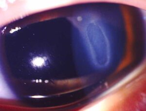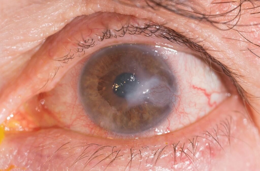
Corneal ulcer healing follows a predictable sequence when the antimicrobial choice is correct and the patient uses the drops.
First, the infiltrate stops growing and its edges blunt, then the epithelium closes over the stroma, and a scar slowly remodels over months. Therefore, each follow-up visit should track these steps in order.
Stages of Corneal Ulcer Healing
Healing begins with control of the organism and the inflammation it provokes, and next, epithelial cells slide in from the wound edge and cover the defect. Keratocytes then transform into fibroblasts and lay down new collagen. As a result, the final scar never matches the transparency or regularity of the original stroma.
The First 48 Hours
The first two days of therapy rarely show dramatic change, and in fact the eye may look slightly worse because inflammation lags behind microbial killing. However, pain usually eases first, and patients often notice it before the examiner sees change. Therefore, judge early response on several signs together and avoid switching drugs after one visit.
All-fit Slit-Lamp Adapter
Record and share exactly what you see at the slit lamp. One adapter fits any slit lamp or surgical microscope — and any smartphone.
From Choroida — the team behind this siteSigns of Improvement
Record the infiltrate and epithelial defect in two perpendicular diameters at every visit. The following findings suggest that treatment is working:
- Blunting of the infiltrate borders, with reduced density
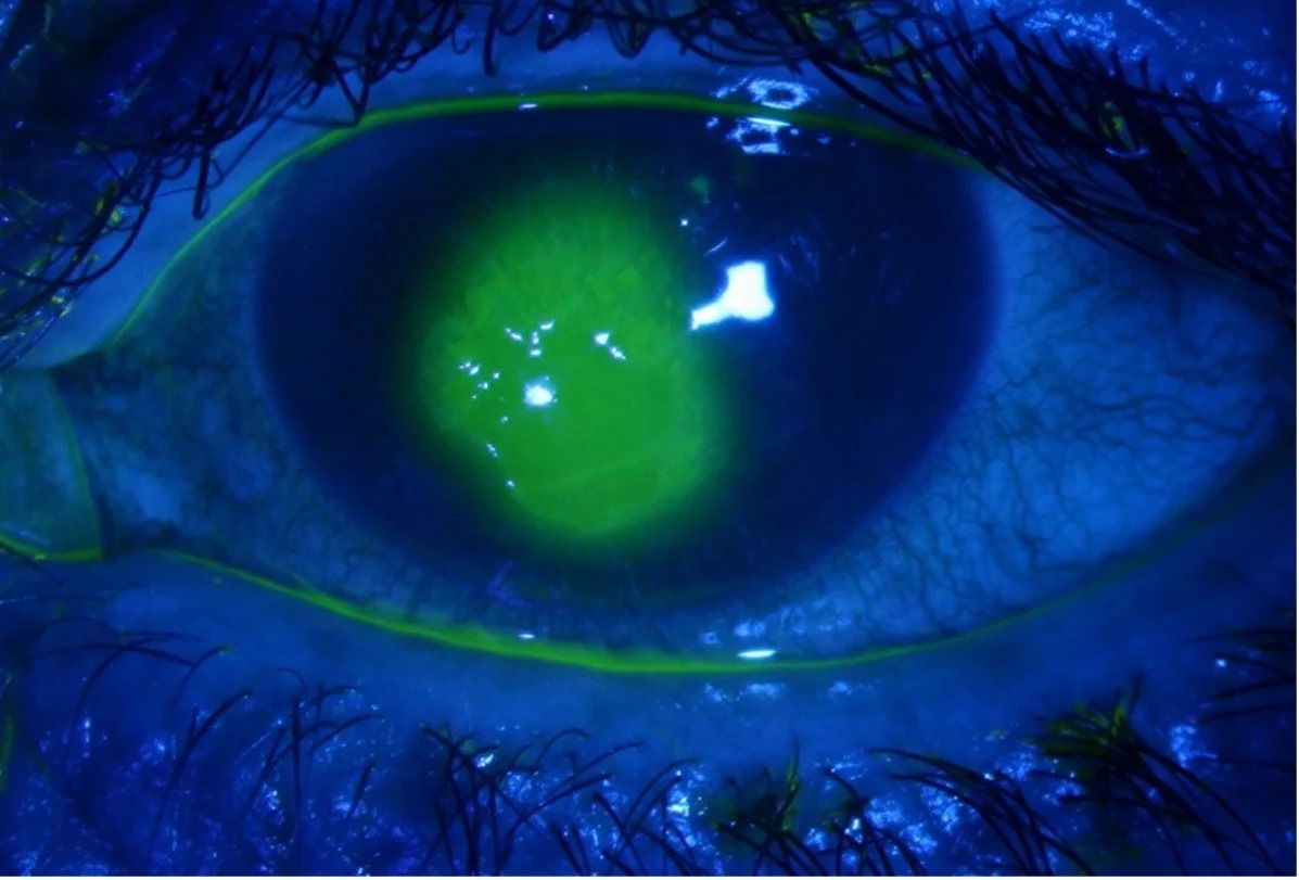
- Shrinking epithelial defect on fluorescein staining
- Less stromal edema and fewer anterior chamber cells
- A smaller or resolving hypopyon
- Reduced pain, discharge and lid swelling
Warning Signs in Corneal Ulcer Healing
Some ulcers progress despite intensive drops, and an enlarging infiltrate, new satellite lesions or a rising hypopyon all signal treatment failure. In addition, progressive stromal thinning raises the risk of descemetocele and perforation. Then confirm adherence, repeat cultures and consider biopsy (see stromal corneal ulcers).
Epithelial Closure in Corneal Ulcer Healing
Closure of the epithelial defect is the clearest milestone, but a persistent defect does not always mean active infection. Frequent fortified drops and preservatives can injure the epithelium and stall closure. As a result, consider tapering toxic medications once the infiltrate is clearly consolidating.
Typical Recovery Time
The pace depends mainly on the organism, ulcer size, depth and location. For example, a small peripheral bacterial ulcer often re-epithelializes within one to two weeks. Fungal and Acanthamoeba keratitis often take weeks to months, and relapse can occur during the taper.
Corneal Ulcer Healing by Organism
Bacterial ulcers usually respond quickly once the correct antibiotic reaches the stroma. In contrast, fungal ulcers respond slowly, and natamycin outperformed voriconazole in the MUTT trial, particularly for Fusarium. Herpetic stromal disease may smolder and recur after apparent quiescence.
Steroids and the Healing Timeline
Topical steroids may reduce scarring once the organism is controlled. However, the SCUT trial found no overall benefit in spectacle-corrected acuity at three months (see the SCUT trial), and most clinicians also avoid steroids in fungal keratitis.
Follow-Up Schedule
Severe ulcers often need daily review until the infiltrate stops progressing. Then visits can extend to every two to three days, and later to weekly. Finally, continue visits through the taper, because recurrence often appears when drop frequency falls.
Scar Formation in Corneal Ulcer Healing
Once the stroma heals, a scar replaces the infiltrate, and its density ranges from faint nebula to dense leucoma (see corneal scar types). Early scars often look denser than their final form, so avoid a final prognosis in the first weeks.
Scar Remodeling Over Months
Stromal remodeling continues for many months after epithelial closure, and as a result haze can fade and vision may improve slowly. However, central scars and irregular astigmatism often limit final acuity. Rigid gas permeable or scleral lenses can mask surface irregularity and help many patients.
Visual Recovery After Corneal Ulcer Healing
Vision depends on scar location more than ulcer size, and a dense peripheral scar, for example, may leave acuity intact. In contrast, a faint central scar can blur vision and cause glare. Also repeat refraction once the surface is stable, since astigmatism often changes.
When Surgery Is Needed
Impending or actual perforation may need tissue adhesive, a patch graft or therapeutic keratoplasty. Later, a dense central scar may justify optical keratoplasty.
Counseling Patients About Corneal Ulcer Healing
Explain early that recovery takes weeks and that vision lags behind symptom relief, and also warn that stopping drops early is a common cause of relapse. In addition, ask patients to return immediately for worsening pain, reduced vision or a new white spot.
Pitfalls to Avoid
- Do not switch antibiotics after one visit because of mild early worsening
- Never confuse drug toxicity with active infection
- Avoid predicting final vision from an early, dense scar
- Do not stop follow-up when the epithelium closes
- Never start steroids before the organism is controlled


Document what you see
Two smartphone imaging tools built for everyday clinic use — one for the slit lamp, one for the fundus.
From Choroida — the team behind this siteReferences
- Lin A, Rhee MK, Akpek EK, et al. Bacterial keratitis Preferred Practice Pattern. Ophthalmology. 2019;126:P1-P55.
- Srinivasan M, Mascarenhas J, Rajaraman R, et al. Corticosteroids for bacterial keratitis: the Steroids for Corneal Ulcers Trial (SCUT). Arch Ophthalmol. 2012;130:143-150.
- Prajna NV, Krishnan T, Mascarenhas J, et al. The Mycotic Ulcer Treatment Trial: a randomized trial comparing natamycin vs voriconazole. JAMA Ophthalmol. 2013;131:422-429.
- Ljubimov AV, Saghizadeh M. Progress in corneal wound healing. Prog Retin Eye Res. 2015;49:17-45.
- Bowling B. Kanski’s Clinical Ophthalmology: A Systematic Approach. 8th ed. Elsevier; 2016.
Test yourself
A few questions straight from this article.
-
In corneal ulcer healing under correct antimicrobial therapy, what is the expected order of events?
The infiltrate first stops growing and its edges blunt, the epithelium then closes over the stroma, and the scar slowly remodels over months; each visit should track these steps in order. -
During stromal repair in corneal ulcer healing, which cells lay down the new collagen?
Keratocytes transform into fibroblasts and lay down new collagen, which is why the final scar never matches the transparency or regularity of the original stroma. -
Why may a corneal ulcer look slightly worse during the first 48 hours of appropriate therapy?
Inflammation lags behind microbial killing, so early change is modest; pain often eases first, and drugs should not be switched after one visit. -
How should the infiltrate and epithelial defect of a corneal ulcer be recorded at each follow-up visit?
Recording both the infiltrate and the epithelial defect in two perpendicular diameters at every visit allows real progress or failure to be tracked. -
Which finding during treatment of a corneal ulcer signals treatment failure?
An enlarging infiltrate, new satellite lesions or a rising hypopyon signal failure, prompting a check on adherence, repeat cultures and consideration of biopsy. -
A corneal ulcer's infiltrate is consolidating but the epithelial defect persists on fortified drops. What is the likely cause?
A persistent defect does not always mean active infection; frequent fortified drops and preservatives can injure the epithelium, so toxic medications may be tapered once the infiltrate consolidates. -
How long does a small peripheral bacterial corneal ulcer typically take to re-epithelialize?
A small peripheral bacterial ulcer often re-epithelializes within one to two weeks, whereas fungal and Acanthamoeba keratitis often take weeks to months. -
In the MUTT trial on fungal corneal ulcers, which drug outperformed voriconazole, particularly for Fusarium?
Fungal ulcers respond slowly, and natamycin outperformed voriconazole in the MUTT trial, particularly for Fusarium. -
What did the SCUT trial find regarding topical steroids for bacterial corneal ulcers?
Steroids may reduce scarring once the organism is controlled, but SCUT found no overall benefit in spectacle-corrected acuity at three months. -
After corneal ulcer healing, which factor most determines final visual acuity?
Vision depends on scar location more than ulcer size: a dense peripheral scar may leave acuity intact, while a faint central scar can blur vision and cause glare.

Writes and reviews the clinical articles on Educate. Every post is checked against peer-reviewed sources before it goes live.