Fungal keratitis is a sight-threatening corneal infection that often looks like a bacterial ulcer at first.
It is common in warm climates and after plant injury.
Early recognition and the right antifungal change the outcome.
Who Gets Fungal Keratitis
Farmers and outdoor workers have the highest risk.
Contact lens wearers and patients on topical steroids also develop it.
A history of vegetable matter injury is classic.
Therefore, always ask about the exact mechanism.
Causative Organisms
Filamentous fungi such as Fusarium and Aspergillus dominate in warm regions.
Candida causes disease in eyes with a compromised surface.
The species affects the response to treatment.
So culture matters whenever it is possible.
All-fit Slit-Lamp Adapter
Record and share exactly what you see at the slit lamp. One adapter fits any slit lamp or surgical microscope — and any smartphone.
From Choroida — the team behind this siteClinical Signs of Fungal Keratitis
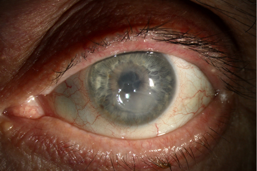
The ulcer has feathery, irregular edges and a dry, raised surface.
Satellite lesions may surround the main infiltrate.
A hypopyon and an endothelial plaque suggest deeper spread.
However, early lesions can look just like bacterial disease (see fungal versus bacterial keratitis).
Symptoms
Pain, redness and blurred vision develop over days.
The course may be slower than in bacterial disease.
Pain can be out of proportion to the signs in some cases.
Also ask about prior treatment with steroids.
Corneal Scraping in Fungal Keratitis
Scrape the base and the edge of the ulcer under a slit lamp.
Place material on a slide for stains and on media for culture.
Potassium hydroxide and Gram stains give rapid answers.
Therefore, scrape before you start drops when possible.
Culture and Confocal Microscopy
Sabouraud agar supports growth of most fungi.
Growth can take days to weeks.
In vivo confocal microscopy shows hyphae directly.
However, availability is limited.
First-Line Fungal Keratitis Treatment
Natamycin 5% is the standard drug for filamentous disease.
Give it hourly while awake for the first days, then taper.
Voriconazole 1% is an alternative for resistant or yeast infections.
Therefore, tailor the drug to the organism.
Evidence From the MUTT Trials
In the Mycotic Ulcer Treatment Trial, natamycin gave better visual outcomes than voriconazole.
Fewer eyes perforated with natamycin.
A second trial added oral voriconazole to topical therapy.
That trial did not show a significant benefit.
Steroids in Fungal Keratitis
Avoid steroids until fungal infection is excluded.
They can worsen the course and enable invasion.
Do not extrapolate trial data from bacterial ulcers (see SCUT).
So keep steroids away from suspected fungal ulcers.
Deep and Recalcitrant Disease
Deep lesions respond poorly to drops alone.
Intrastromal or intracameral injections are options in selected eyes.
Collagen cross-linking has shown mixed results.
Therefore, refer early when the infiltrate is deep.
Surgical Treatment
Therapeutic keratoplasty is needed for perforation or uncontrolled infection.
Early graft improves the chance of saving the eye.
Delayed surgery risks spread into the anterior chamber.
So plan for surgery if the ulcer worsens.
Follow-Up
Review daily at first and then at wider intervals.
Look for a smaller infiltrate, less pain and a healing epithelium.
Treatment may continue for weeks.
Also watch for secondary glaucoma.
Counseling Patients
Explain that healing is slow and scarring is common.
Stress the need to complete treatment.
Advise eye protection for farm work.
Also warn contact lens wearers about the risk.
Pitfalls to Avoid
- Do not start steroids before excluding fungus
- Never rely on appearance alone to separate fungal from bacterial disease
- Avoid stopping antifungals too early
- Do not delay referral of deep ulcers
Fungal Keratitis in Contact Lens Wearers
Fungal keratitis is less common than bacterial disease in lens wearers.
However, certain solutions were linked to Fusarium outbreaks in the past.
Therefore, ask about the product and stop lens wear at once.
Also culture the case and solution when possible.
Fungal Keratitis After Plant Trauma
Branch, thorn and crop injuries introduce fungi into the stroma.
Symptoms may start days after a trivial injury.
Because the history is easy to forget, ask directly about plants.
Then scrape every suspicious ulcer.
Fungal Keratitis and Steroid Use
Prior steroid treatment can mask early signs.
If an ulcer worsens on steroids, suspect fungus.
Then stop the steroid and scrape the cornea.
As a result, the correct drug can start without delay.
Counseling About Fungal Keratitis
Because treatment lasts weeks, explain the course at the first visit.
Patients should know that healing is slow even when the drug is right.
If the eye gets worse, they should call at once.
In addition, remind farm workers to wear eye protection.
Fungal Keratitis Prevention
Safety glasses lower the risk of plant injury.
When an injury occurs, patients should seek care the same day.
Also treat chronic surface disease before it invites infection.
Therefore, prevention begins with education.
For the latest studies, see the PubMed results for fungal keratitis.


Document what you see
Two smartphone imaging tools built for everyday clinic use — one for the slit lamp, one for the fundus.
From Choroida — the team behind this siteReferences
- Prajna NV, Krishnan T, Mascarenhas J, et al. The Mycotic Ulcer Treatment Trial: a randomized trial comparing natamycin vs voriconazole. JAMA Ophthalmol. 2013;131:422-429.
- Prajna NV, Krishnan T, Rajaraman R, et al. Effect of oral voriconazole on fungal keratitis in the Mycotic Ulcer Treatment Trial II. JAMA Ophthalmol. 2016;134:1365-1372.
- Thomas PA, Kaliamurthy J. Mycotic keratitis: epidemiology, diagnosis and management. Clin Microbiol Infect. 2013;19:210-220.
- Brown L, Leck AK, Gichangi M, Burton MJ, Denning DW. The global incidence and diagnosis of fungal keratitis. Lancet Infect Dis. 2021;21:e49-e57.
Test yourself
A few questions straight from this article.
-
Which injury history is classic for fungal keratitis?
Farmers and outdoor workers have the highest risk, and branch, thorn or crop injury introduces fungi into the stroma, so the exact mechanism should always be asked. -
Which organism causes fungal keratitis mainly in eyes with a compromised ocular surface?
Filamentous fungi such as Fusarium and Aspergillus dominate in warm regions, while Candida causes disease in eyes with a compromised surface. -
Which corneal ulcer appearance suggests fungal keratitis?
A fungal ulcer has feathery, irregular edges and a dry raised surface, often with satellite lesions, though early lesions can look just like bacterial disease. -
Which stain gives a rapid answer on corneal scrapings from suspected fungal keratitis?
Scraping the base and edge of the ulcer for potassium hydroxide and Gram stains gives rapid answers, ideally before drops are started. -
Which test shows fungal hyphae directly in the living cornea?
In vivo confocal microscopy shows hyphae directly but availability is limited; culture on Sabouraud agar can take days to weeks. -
What is the standard first-line drug for filamentous fungal keratitis?
Natamycin 5% is standard for filamentous disease, given hourly while awake at first and then tapered; voriconazole 1% is an alternative for resistant or yeast infections. -
In the Mycotic Ulcer Treatment Trial, how did natamycin compare with voriconazole for fungal keratitis?
Natamycin gave better visual outcomes than voriconazole, and fewer eyes perforated; a second trial adding oral voriconazole showed no significant benefit. -
A corneal ulcer worsens while the patient is on topical steroids; what should be suspected and done?
Steroids can mask early signs and enable fungal invasion, so an ulcer worsening on steroids suggests fungus: stop the steroid and scrape so the right drug starts without delay. -
When is therapeutic keratoplasty needed in fungal keratitis?
Therapeutic keratoplasty is needed for perforation or uncontrolled infection; early grafting improves the chance of saving the eye, while delay risks spread into the anterior chamber. -
For a deep fungal corneal infiltrate responding poorly to drops alone, which treatment option is used in selected eyes?
Deep lesions respond poorly to drops alone, so intrastromal or intracameral injections are options in selected eyes, and deep infiltrates warrant early referral.

Writes and reviews the clinical articles on Educate. Every post is checked against peer-reviewed sources before it goes live.