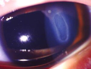
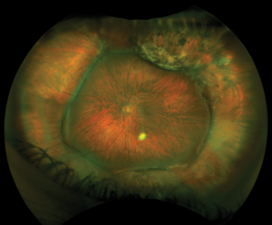
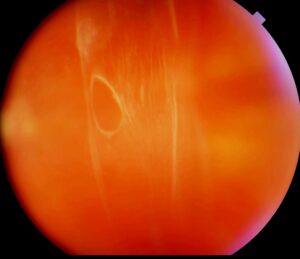
Cataract surgery in diabetic patients works well but carries a higher risk of macular problems.
Surgery can trigger inflammation that worsens edema in eyes with retinopathy, so planning before and after surgery protects the macula and the final vision.
Why Diabetic Eyes Need Extra Care
Diabetes breaks down the blood-retinal barrier, and surgical inflammation adds a second hit to the same barrier. Cataract also often develops earlier and faster in diabetes, so many diabetic patients need surgery and face this risk.
Retinopathy Progression After Cataract Surgery
Older reports suggested that retinopathy worsens after cataract surgery, but modern studies with small incisions show a smaller effect. Squirrell and colleagues also found limited progression after uncomplicated phacoemulsification in type 2 diabetes, so risk depends on the stage of disease and the quality of surgery.
Fundus Explorer Pro
Photograph the retinal findings described here with the phone already in your pocket — 22 D optics and built-in illumination in one handheld unit.
From Choroida — the team behind this siteMacular Edema After Cataract Surgery
Pseudophakic macular edema is more common in diabetic eyes, and Chu and colleagues found that the risk rose with the severity of retinopathy. Eyes with edema before surgery are at the highest risk, so assess the macula first.
Cataract Surgery in Diabetic Patients: Preoperative Assessment
Grade the retinopathy with a dilated examination, and perform OCT of the macula in every diabetic patient. Also check for neovascularization of the iris and angle, and review glycemic control and blood pressure.
Treating Edema Before Cataract Surgery
Center-involving edema should be treated before surgery when possible, and anti-VEGF injections or laser reduce the swelling (see diabetic macular edema). Delaying surgery by a few weeks is reasonable if vision allows, so a dry macula gives a safer start.
Treating Proliferative Disease Before Cataract Surgery
Active new vessels need treatment before surgery, and panretinal photocoagulation lowers the risk of progression (see panretinal photocoagulation). Media opacity may block laser, so treat early and plan the timing with the retina team.
Cataract Surgery Technique in Diabetes
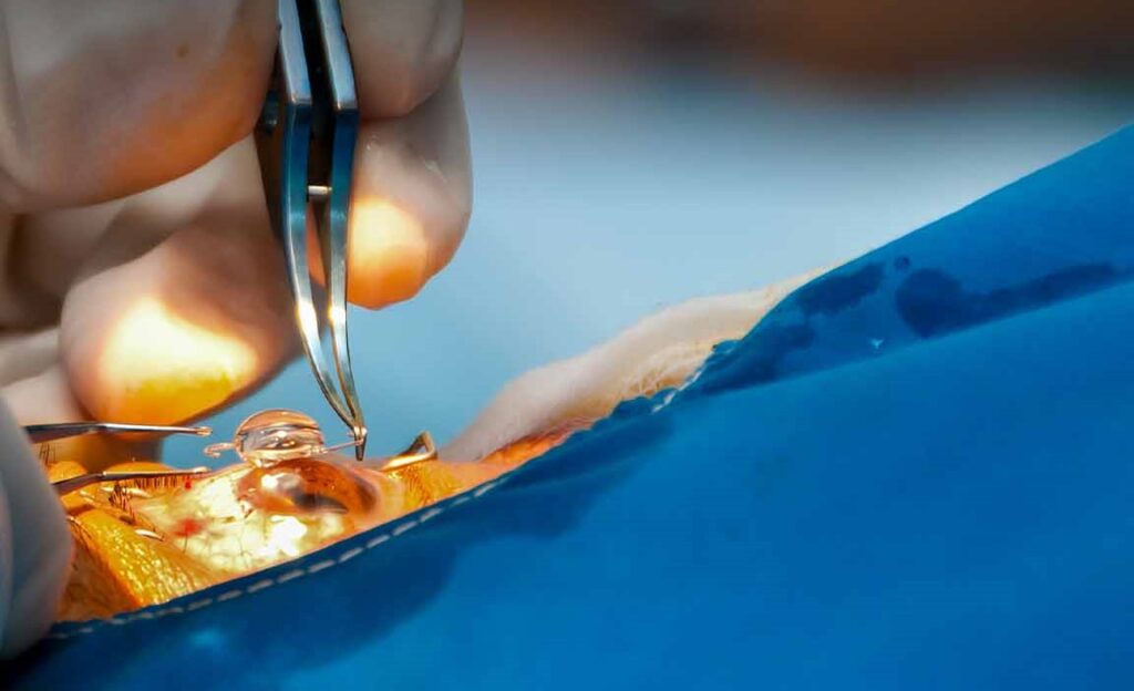
Use a clear corneal incision of small size and minimal trauma, and create a well-centered capsulorhexis and a clean capsule. Also avoid iris manipulation and long phaco time, so gentle surgery reduces inflammation.
Intraocular Lens Choice
A monofocal lens suits most diabetic eyes, while multifocal lenses reduce contrast and can complicate retinal care. Large optics also help with future fundus exams, so avoid lenses that block the view of the periphery.
Anti-Inflammatory Prophylaxis
Combine a topical steroid with a nonsteroidal drop in diabetic patients. The ESCRS PREMED study tested prophylaxis in diabetic eyes, and the combination lowered macular thickening compared with steroid alone, so many surgeons use both agents for several weeks.
Intravitreal Therapy at Surgery
Some surgeons inject anti-VEGF drugs or steroids at the end of surgery in high-risk eyes, but the evidence for routine use is mixed. DRCR studies also address eyes with edema before surgery, so use it for selected cases.
Follow-Up After Cataract Surgery
Examine the patient at one day, one week and one month, and perform OCT at one month in eyes with retinopathy. Also look for iris new vessels and pressure rise, so early detection allows prompt treatment.
Managing Postoperative Edema
Start or intensify nonsteroidal drops and steroids, and add anti-VEGF injections or a steroid implant if edema persists. Dowler and colleagues showed that some edema resolves spontaneously, but treat sooner if vision is poor.
Glycemic Control
Poor control raises the risk of complications, and rapid improvement before surgery may transiently worsen retinopathy. Aim for steady control over weeks, so coordinate with the diabetes team.
Counseling Patients
Explain that vision may improve less than in nondiabetic patients, and warn that edema can appear after surgery and needs treatment. Remind patients about regular retinal visits, so expectations stay realistic.
Pitfalls to Avoid
- Do not operate on an eye with untreated center-involving edema without a plan
- Never skip a macular OCT before surgery
- Do not forget the postoperative OCT in eyes with retinopathy
- Never ignore early iris new vessels
Intraoperative Complications
Posterior capsule tear and vitreous loss raise the risk of macular edema, and small pupils in diabetes complicate surgery. Use pupil expansion devices as needed, so plan for difficult pupils.
Role of Retinal Specialists
Co-manage patients with severe retinopathy, and agree on the timing of laser, injections and surgery. Share the results of OCT, so care stays coordinated.


Document what you see
Two smartphone imaging tools built for everyday clinic use — one for the slit lamp, one for the fundus.
From Choroida — the team behind this siteReferences
- Chu CJ, Johnston RL, Buscombe C, et al. Risk factors and incidence of macular edema after cataract surgery: a database study of 81984 eyes. Ophthalmology. 2016;123:316-323.
- Squirrell D, Bhola R, Bush J, Winder S, Talbot JF. A prospective, case controlled study of the natural history of diabetic retinopathy and maculopathy after uncomplicated phacoemulsification cataract surgery in patients with type 2 diabetes. Br J Ophthalmol. 2002;86:565-571.
- Dowler JG, Sehmi KS, Hykin PG, Hamilton AM. The natural history of macular edema after cataract surgery in diabetes. Ophthalmology. 1999;106:663-668.
- Wielders LHP, Schouten JSAG, Winkens B, et al. ESCRS PREMED study report 2: results of a European multicentre trial of the prevention of cystoid macular edema after cataract surgery in diabetics. J Cataract Refract Surg. 2018;44:836-847.
Test yourself
A few questions straight from this article.
-
Why are diabetic eyes more prone to macular edema after cataract surgery?
Diabetes already breaks down the blood-retinal barrier, and surgical inflammation delivers a second hit to the same barrier, worsening edema in eyes with retinopathy. -
With modern small-incision cataract surgery, how much does diabetic retinopathy progress compared with older reports?
Older reports suggested retinopathy worsens after cataract surgery, but small-incision studies show a smaller effect, with risk depending on disease stage and surgical quality. -
Which diabetic eyes face the highest risk of macular edema after cataract surgery?
Pseudophakic macular edema risk rises with retinopathy severity, and eyes that already have edema before surgery carry the highest risk, so the macula is assessed first. -
Which macular test should every diabetic patient have before cataract surgery?
Preoperative assessment includes a dilated examination to grade retinopathy and a macular OCT in every diabetic patient, plus a check for iris and angle neovascularization. -
How should center-involving diabetic macular edema be handled when cataract surgery is planned?
Center-involving edema should be treated before surgery when possible; delaying surgery a few weeks is reasonable if vision allows, because a dry macula gives a safer start. -
In a diabetic patient with active proliferative retinopathy and a cataract, why should panretinal photocoagulation be done early?
Active new vessels need treatment before surgery to lower the risk of progression, and because media opacity may block laser, treatment is timed early with the retina team. -
Which intraocular lens type suits most diabetic eyes undergoing cataract surgery?
A monofocal lens suits most diabetic eyes, since multifocal lenses reduce contrast and can complicate retinal care, and large optics help future peripheral fundus examination. -
Which prophylaxis lowered macular thickening in diabetic eyes after cataract surgery in the ESCRS PREMED study?
In the ESCRS PREMED study, combining a topical steroid with a nonsteroidal drop lowered macular thickening compared with steroid alone, so many surgeons use both agents. -
After cataract surgery in a diabetic eye with retinopathy, when is a macular OCT recommended?
Patients are examined at one day, one week and one month, with OCT at one month in eyes with retinopathy, while also watching for iris new vessels and pressure rise. -
Why should glycemic control be improved steadily rather than rapidly before cataract surgery in diabetes?
Poor control raises complication risk, but rapid improvement before surgery may transiently worsen retinopathy, so steady control over weeks is coordinated with the diabetes team.

Writes and reviews the clinical articles on Educate. Every post is checked against peer-reviewed sources before it goes live.