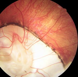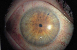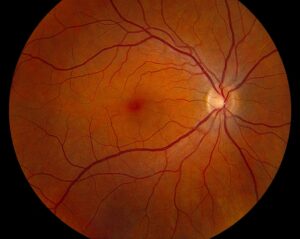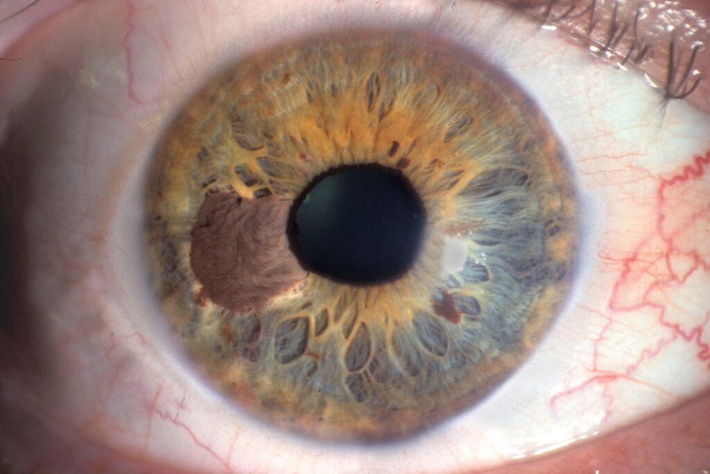
Uveal melanoma surveillance aims to find liver metastases early, because about half of patients eventually develop them.
The eye tumor is treated successfully in most cases, but cells may already have spread through the blood.
Therefore, follow-up must continue long after the eye is stable.
Why Uveal Melanoma Surveillance Matters
The Collaborative Ocular Melanoma Study found that the liver is the first and dominant site of spread.
Kujala and colleagues showed that deaths continue to occur beyond 10 years.
As a result, a clear eye does not end the risk (see choroidal melanoma).
Risk Stratification in Uveal Melanoma Surveillance
- Tumor size, ciliary body involvement, and extraocular extension
- Monosomy 3 and chromosome 8q gain
- Gene expression class 2, as reported by Onken and colleagues
Larger tumors and class 2 profiles carry the highest risk, so they merit the closest follow-up.
Also, a class 1A profile predicts a low risk of metastasis.
Fundus Explorer Pro
Photograph the retinal findings described here with the phone already in your pocket — 22 D optics and built-in illumination in one handheld unit.
From Choroida — the team behind this siteImaging for Uveal Melanoma Surveillance
Most centers use liver imaging with ultrasound or magnetic resonance imaging every 6 to 12 months.
Magnetic resonance imaging is more sensitive than ultrasound for small lesions.
Also, liver function tests are cheap, but they miss small metastases.
No schedule has been proven to improve survival, so protocols differ between centers (see plaque brachytherapy).
What Early Detection Offers in Uveal Melanoma Surveillance
Rantala and colleagues showed that patients with a small liver burden live longer after surgery or liver-directed therapy.
However, selection bias may explain part of this effect.
Still, early detection allows eligibility for trials and for new systemic treatment.
Counselling Patients on Uveal Melanoma Surveillance
Explain the plan in plain terms and avoid frightening statistics.
Also tell patients that surveillance protects them, because early detection widens the options.
Many patients feel anxious before each scan, so schedule results visits promptly.
Give a written plan that names who orders the imaging and who receives the result.
Coordinating Care for Uveal Melanoma Surveillance
The ocular oncologist usually sets the schedule, and a medical oncologist or hepatologist reviews the images.
Therefore, document the tumor class and the next scan date in the chart.
When imaging shows a lesion, refer to a center with a multidisciplinary team for biopsy and treatment planning.
Finally, remember that second primary cancers occur, so general health screening still matters.
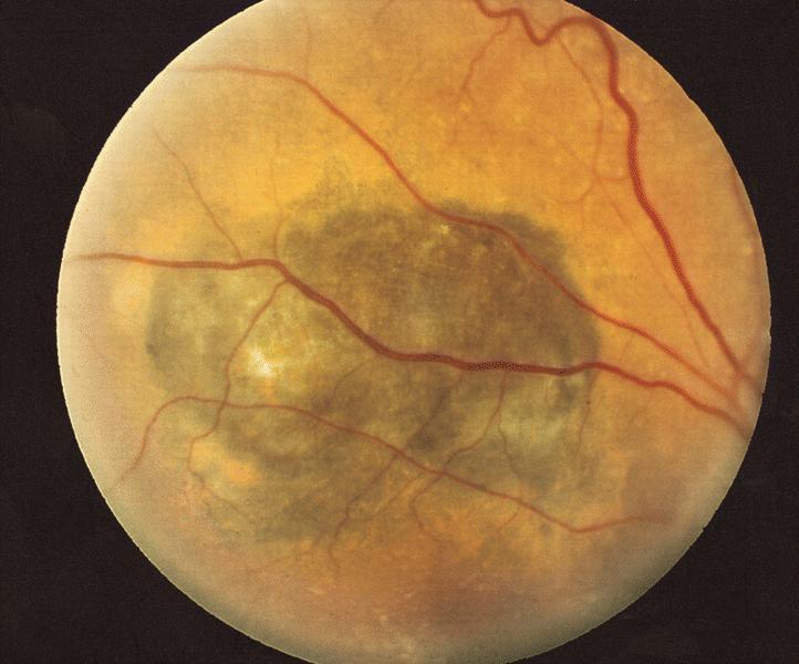
Prognostic Testing in Uveal Melanoma
Jager and colleagues reviewed the genetic features that predict spread.
Loss of one copy of chromosome 3 and BAP1 mutation carry a high risk, while SF3B1 and EIF1AX mutations carry lower risk.
Also, a gene expression profile divides tumors into class 1 and class 2.
Therefore, ask for testing after biopsy or enucleation, and use the result to set the interval.
Beyond the Liver in Uveal Melanoma Surveillance
The liver is the first site in most patients, but lung, bone, and skin metastases occur.
Therefore, symptoms such as cough, bone pain, or skin nodules need prompt evaluation.
Also, consider whole-body imaging when liver imaging shows disease or when symptoms point elsewhere.
Because follow-up can last for decades, explain the plan again at each annual visit.
Practical Points for the Clinician
Keep a simple register of patients with uveal melanoma, and record the date of the last liver scan.
Also set a recall system, because patients lost to follow-up are common.
Review any new symptoms of weight loss, abdominal pain, or fatigue at each visit.
Then copy the primary care physician on the surveillance plan.
Open Questions in Surveillance
Researchers still debate how often to scan and which modality is best.
Also, circulating tumor DNA and liquid biopsies are under study as screening tools.
Therefore, expect protocols to change as evidence grows.
Key Points for the Clinician
Metastasis most often appears in the liver, and it can appear many years after treatment.
Therefore, keep patients under follow-up for life.
Also, stratify risk by genetics and clinical features, and match the scan interval to the risk.
Finally, coordinate with oncology so that no patient falls between services.
Because evidence on schedules is limited, document the reasoning for the plan.
Supporting the Patient
Many patients live with the fear of recurrence for years.
Also offer access to psychological support and patient groups.
Because uncertainty is hard, give clear dates for the next scan.


Document what you see
Two smartphone imaging tools built for everyday clinic use — one for the slit lamp, one for the fundus.
From Choroida — the team behind this siteReferences
- Collaborative Ocular Melanoma Study Group. Assessment of metastatic disease status at death in 435 patients with large choroidal melanoma in COMS report no. 15. Arch Ophthalmol. 2001;119:670-676.
- Kujala E, Makitie T, Kivela T. Very long-term prognosis of patients with malignant uveal melanoma. Invest Ophthalmol Vis Sci. 2003;44:4651-4659.
- Onken MD, Worley LA, Char DH, et al. Collaborative Ocular Oncology Group report number 1: prospective validation of a multi-gene prognostic assay in uveal melanoma. Ophthalmology. 2012;119:1596-1603.
- Rantala ES, Hernberg M, Kivela TT. Overall survival after treatment for metastatic uveal melanoma: a systematic review and meta-analysis. Melanoma Res. 2019;29:561-568.
- Jager MJ, Shields CL, Cebulla CM, et al. Uveal melanoma. Nat Rev Dis Primers. 2020;6:24.
Test yourself
A few questions straight from this article.
-
Roughly what proportion of patients with uveal melanoma eventually develop liver metastases?
About half of patients eventually develop liver metastases, because cells may already have spread through the blood even when the eye tumor is treated successfully. -
According to the Collaborative Ocular Melanoma Study, which organ is the first and dominant site of uveal melanoma spread?
COMS found the liver to be the first and dominant site of spread, which is why surveillance centres on liver imaging. -
Why should patients treated for uveal melanoma stay under surveillance for life?
Deaths continue to occur beyond 10 years and metastasis can appear many years after treatment, so a clear eye does not end the risk. -
Which uveal melanoma gene expression profile class carries the highest metastatic risk and merits the closest follow-up?
Larger tumors and class 2 profiles carry the highest risk, whereas a class 1A profile predicts a low risk of metastasis. -
Which genetic feature of uveal melanoma carries a high risk of metastasis?
Loss of one copy of chromosome 3 and BAP1 mutation carry a high risk of spread, while SF3B1 and EIF1AX mutations carry lower risk. -
At what interval do most centers image the liver during uveal melanoma surveillance?
Most centers use liver ultrasound or MRI every 6 to 12 months, matching the interval to the patient's risk. -
Which liver imaging modality is more sensitive for small uveal melanoma metastases?
MRI is more sensitive than ultrasound for small liver lesions, although both are used for surveillance. -
What is the main weakness of liver function tests in uveal melanoma surveillance?
Liver function tests are cheap, but they miss small metastases, so imaging remains the basis of surveillance. -
Why do uveal melanoma surveillance protocols differ between centers?
No surveillance schedule has been proven to improve survival, so protocols vary and the reasoning for each plan should be documented. -
What should happen when surveillance imaging shows a liver lesion in a patient with uveal melanoma?
A lesion on imaging should prompt referral to a center with a multidisciplinary team for biopsy and treatment planning; early detection widens options, including trials.

Writes and reviews the clinical articles on Educate. Every post is checked against peer-reviewed sources before it goes live.