
The anterior segment of the eye gets most of its blood from the anterior ciliary arteries, which travel with the rectus muscles.
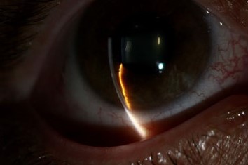
When several of these muscles are detached in the same eye, the blood supply may fall below what the cornea, iris, and ciliary body need.
The result is anterior segment ischemia (ASI), a rare but potentially serious complication.
How it happens
Each rectus muscle carries two anterior ciliary arteries, except the lateral rectus, which carries one.
These arteries join the major arterial circle of the iris, and they supply the ciliary body, iris, and part of the limbal and conjunctival circulation.
The posterior ciliary arteries provide the rest, mostly to the posterior segment.
If the anterior ciliary arteries are cut or occluded during surgery, the blood flow to the anterior segment falls.
In a healthy eye with intact collateral circulation, removal of one or two muscles is usually tolerated, and problems occur when more muscles are involved.
Risk factors
- Surgery on three or more rectus muscles in the same eye at one time, and particularly operations on both vertical rectus muscles together with a horizontal one
- Previous muscle surgery or scleral buckle, which have already reduced the blood supply
- Older age and vascular disease such as carotid stenosis, diabetes, sickle cell disease, and giant cell arteritis
- Thyroid eye disease with tight muscles
- Extensive conjunctival dissection and cautery near the limbus
All-fit Slit-Lamp Adapter
Record and share exactly what you see at the slit lamp. One adapter fits any slit lamp or surgical microscope — and any smartphone.
From Choroida — the team behind this sitePresentation
Symptoms usually appear within the first few days, but may be delayed.
- Pain, redness, and photophobia
- Blurred vision
- Corneal edema and Descemet folds
- Anterior chamber cells and flare, which can be marked
- Iris changes: sectoral or diffuse atrophy, a dilated, poorly reactive or distorted pupil
- Low intraocular pressure from decreased aqueous production, and sometimes cataract and phthisis in severe cases
Careful examination, including iris angiography when available, may show filling defects in the iris.
Differential diagnosis
Postoperative inflammation, endophthalmitis, and toxic reactions can look similar.
ASI tends to produce a deep, quiet inflammation with signs of ischemia, such as iris atrophy and low pressure, and the clinician should consider it whenever an eye that has had extensive muscle surgery becomes inflamed.
Management
- Corticosteroids topical and sometimes systemic, to limit inflammation and protect tissues
- Cycloplegics for comfort and to prevent synechiae
- Careful monitoring for infection, secondary glaucoma, and complications
- In some cases, the effect of ischemia is temporary and the circulation recovers through collaterals
- Evaluate for systemic vascular disease that increased the risk
Prevention
- Avoid surgery on more than two rectus muscles in one eye at one session, particularly in older patients and those with vascular disease. Stage surgery, leaving at least six months between procedures, when three or more muscles need treatment
- Use techniques that spare the anterior ciliary vessels where possible, such as vessel-sparing muscle surgery
- Weigh the risks in patients with sickle cell disease, thyroid eye disease, and prior surgery
- Discuss the risk with the patient at the time of consent
Early signs worth looking for
In the first days after strabismus surgery, a very mild form may show only a slight reduction in intraocular pressure, mild cells and flare, and a slightly sluggish pupil. These signs are easy to attribute to ordinary postoperative inflammation. Be suspicious if the pain is greater than expected, the cornea is hazy, or the iris looks dull. Ask whether more than two rectus muscles were operated on, and whether there were previous operations.
Testing and imaging
Fluorescein or indocyanine green iris angiography can show filling defects, which confirm the diagnosis in uncertain cases. Anterior segment OCT may show corneal thickening and Descemet folds. Check the pressure often, since hypotony may lead to choroidal effusion and macular folds in severe cases.
Counselling and consent
Before surgery on several muscles, explain that there is a small risk of reduced blood supply to the front of the eye, and describe the steps taken to avoid it, such as staging the procedures. After surgery, instruct the patient to report pain or visual change promptly. In older patients with vascular disease, the surgeon should consider alternative approaches, such as the use of botulinum toxin or adjustable procedures that spare vessels.
Differential diagnosis in the first week
Other causes of a painful, inflamed eye after strabismus surgery include infection, scleral perforation, allergy to materials or drops, and a retained suture reaction. ASI is a diagnosis of exclusion that depends on the surgical history and the pattern of findings, so consider infection first and treat it if there is doubt.
Prognosis
Mild cases recover well.
Severe cases may leave iris atrophy, permanent hypotony, cataract, or phthisis.
The best strategy is prevention through planning.


Document what you see
Two smartphone imaging tools built for everyday clinic use — one for the slit lamp, one for the fundus.
From Choroida — the team behind this siteReferences
- Olver JM, Lee JP. The effects of strabismus surgery on anterior segment circulation. Eye. 1989;3:318-326.
- France TD, Simon JW. Anterior segment ischemia syndrome following muscle surgery: the AAPO&S experience. J Pediatr Ophthalmol Strabismus. 1986;23:87-91.
- Saunders RA, Phillips MS. Anterior segment ischemia after three rectus muscle surgery. Ophthalmology. 1988;95:533-537.
- Kanski JJ, Bowling B. Clinical Ophthalmology: A Systematic Approach. 7th ed. London: Elsevier; 2011.
Test yourself
A few questions straight from this article.
-
Which vessels supply most of the blood to the anterior segment of the eye?
The anterior segment gets most of its blood from the anterior ciliary arteries, which travel with the rectus muscles and join the major arterial circle of the iris. -
How many anterior ciliary arteries does each rectus muscle carry?
Each rectus muscle carries two anterior ciliary arteries, with the exception of the lateral rectus, which carries one. -
Which surgical plan carries the highest risk of anterior segment ischemia?
Removal of one or two muscles is usually tolerated in a healthy eye; risk rises with surgery on three or more rectus muscles at one time, especially both vertical recti plus a horizontal. -
When three or more rectus muscles need surgery, what staging interval is advised between procedures?
Surgery should be staged with at least six months left between procedures when three or more rectus muscles require treatment. -
Why is intraocular pressure low in anterior segment ischemia after rectus muscle surgery?
Low intraocular pressure in anterior segment ischemia results from decreased aqueous production, reflecting compromised perfusion of the ciliary body. -
What does fluorescein or indocyanine green iris angiography show in anterior segment ischemia?
Fluorescein or indocyanine green iris angiography demonstrates filling defects in the iris, which is useful in uncertain cases. -
Why are cycloplegics used in the treatment of anterior segment ischemia?
Cycloplegics are given for comfort and to prevent synechiae, alongside corticosteroids to limit inflammation and careful monitoring for complications. -
In a painful, inflamed eye days after strabismus surgery, which diagnosis should be considered and treated first if there is doubt?
Anterior segment ischemia is a diagnosis of exclusion resting on the surgical history and the pattern of findings, so infection should be considered first and treated if there is doubt. -
Which findings may persist after a severe episode of anterior segment ischemia?
Mild cases recover well, but severe anterior segment ischemia may leave iris atrophy, permanent hypotony, cataract or phthisis. -
Which combination suggests a very mild anterior segment ischemia in the first days after strabismus surgery?
A very mild form may show only slightly reduced intraocular pressure, mild cells and flare and a slightly sluggish pupil, signs easily mistaken for ordinary postoperative inflammation.
