
Pupillary capture is a mechanical problem.
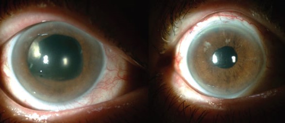
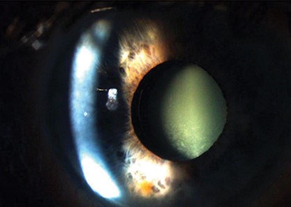
The edge of the pupil slips around the optic of a posterior chamber intraocular lens, so that the iris sits in front of a portion of the lens that should be behind it.
The pupil is displaced, the optic edge may be visible, and the patient complains of glare, halos, or blur.
How it occurs
Normally, an IOL in the capsular bag or ciliary sulcus sits behind the iris, and the pupil margin rests on the anterior surface of the optic.
If the lens is pushed forward, or if the iris moves behind the lens, the pupil may pass over the optic edge and the optic can herniate through the pupil.
The optic is “captured” in front of the iris, partly or completely.
Why it happens
- Contraction or fibrosis of the capsular bag (phimosis), which pushes the optic forward
- Haptic placement in the sulcus of a lens designed for the bag, which tilts the optic
- Posterior capsule rupture or zonular weakness, in which lens support is poor
- A flat anterior chamber or inflammation that changes the position of the iris
- Small pupils and posterior synechiae in uveitis
- Lens designs with a large optic or a particular haptic angulation
- In a pediatric eye, deliberate optic capture is sometimes used as a technique, with the optic passed through the posterior capsulorhexis to stabilize the lens (see Soemmering ring after cataract surgery for a related capsular bag phenomenon)
All-fit Slit-Lamp Adapter
Record and share exactly what you see at the slit lamp. One adapter fits any slit lamp or surgical microscope — and any smartphone.
From Choroida — the team behind this siteClinical features
- Decentration or ovalization of the pupil
- The optic edge crossing the pupillary area, producing glare and ghost images
- Variable vision as the iris shifts with pupil size and head position
- Pigment dispersion, uveitis, or raised pressure if there is chronic iris chafing
- Lens tilt visible at the slit lamp, and sometimes a changed refraction
Diagnosis
The diagnosis is made at the slit lamp.
Examine with the pupil undilated and then dilated.
Anterior segment OCT or ultrasound biomicroscopy helps to define the relationship of the iris, optic, and haptics, and to see whether the lens is in the bag or sulcus (see anterior segment OCT).
Management
- Pharmacological treatment can sometimes release capture: dilating the pupil, followed by supine positioning and a miotic, may free the iris. This is useful in recent cases.
- Surgical repositioning is usually needed when capture is chronic. The optic is released and replaced behind the iris. If there is contraction of the bag, an anterior capsulotomy with relaxing cuts, or YAG capsulotomy, may be required.
- Lens exchange or fixation may be necessary if the zonular support is poor. Sutures or scleral fixation can then be used.
- Treat the cause: control inflammation, deal with synechiae, and manage pressure.
Prevention
Careful capsulorhexis sizing, placement of the lens in the bag whenever possible, and use of a lens suitable for the sulcus when bag fixation is not possible, are the main preventive measures.
In patients with a tendency to bag contraction, a larger capsulorhexis and a capsular tension ring may help.
What the patient notices
Patients may complain of glare, a shadow, an irregular pupil, or fluctuating vision that changes with the lighting or head position. Some notice that the pupil looks different in photographs. In a few cases, the optic is visible through the pupil in front of the iris, and the patient has a cosmetic concern. Pain and redness are unusual, except when chronic chafing produces inflammation.
Differential diagnosis
Pupillary capture should be separated from a tilted or decentered lens, from posterior synechiae with a distorted pupil, from iris atrophy, and from a post-traumatic pupil. A careful slit-lamp examination with the pupil in different sizes will show whether the iris lies in front of or behind the optic. Imaging of the anterior segment is helpful when the diagnosis is uncertain.
Why early correction is better
If the iris is trapped for a long period, it may become atrophic or develop synechiae, and the fibrosis of the capsular bag may be more difficult to deal with. A short course of treatment to release the capture in the first days after surgery, with positioning and pupil manipulation, is often sufficient. Delay can turn a simple problem into one that needs a return to the operating room.
Prevention in daily practice
Surgeons can reduce the risk by sizing the capsulorhexis so that it overlaps the optic edge, by removing viscoelastic carefully behind the lens, and by choosing a lens whose haptic design matches the planned position. In patients with small pupils, synechiae, or an unstable bag, a note in the chart and a plan for early postoperative review pay off.
Prognosis
The outlook is good if the problem is recognized and corrected before chronic complications have developed.


Document what you see
Two smartphone imaging tools built for everyday clinic use — one for the slit lamp, one for the fundus.
From Choroida — the team behind this siteReferences
- Apple DJ, Mamalis N, Loftfield K, et al. Complications of intraocular lenses. A historical and histopathological review. Surv Ophthalmol. 1984;29:1-54.
- Stark WJ, Maumenee AE, Datiles M, et al. Intraocular lenses: complications and visual results. Trans Am Ophthalmol Soc. 1983;81:280-309.
- Gimbel HV. Posterior capsulorhexis with optic capture: maintaining a clear visual axis after pediatric cataract surgery. J Cataract Refract Surg. 1994;20:658-664.
- Davison JA. Capsule contraction syndrome. J Cataract Refract Surg. 1993;19:582-589.
Test yourself
A few questions straight from this article.
-
Pupillary capture of an intraocular lens describes which configuration?
The pupil margin slips around the optic of a posterior chamber lens so the iris sits in front of a portion of the lens that should lie behind it, and the optic is captured anteriorly. -
Which pharmacological sequence can release a recent pupillary capture?
Dilating the pupil, then placing the patient supine and giving a miotic, can free the iris and release the capture; this works best in recent cases. -
Which change most often pushes an intraocular lens optic forward through the pupil?
Contraction or fibrosis of the capsular bag pushes the optic forward so the pupil can pass over its edge; sulcus placement of a bag lens, poor lens support and iris position changes also contribute. -
In which setting is optic capture used deliberately as a surgical technique?
In a pediatric eye the optic is sometimes passed deliberately through the posterior capsulorhexis to stabilise the lens, so capture is used as a technique rather than treated as a complication. -
Which symptom pattern fits pupillary capture of an intraocular lens?
The optic edge crossing the pupillary area produces glare and ghost images, and vision varies as the iris shifts with pupil size and head position; pain and redness are unusual. -
Which imaging defines the relationship of iris, optic and haptics in pupillary capture?
Diagnosis is clinical, at the slit lamp with the pupil undilated and then dilated, and anterior segment OCT or ultrasound biomicroscopy shows whether the lens sits in the bag or the sulcus. -
What does chronic iris chafing against a captured intraocular lens optic produce?
Chronic chafing of the iris against the captured optic causes pigment dispersion, uveitis and raised intraocular pressure, which is why early release is preferred. -
Which examination step separates pupillary capture from a merely tilted or decentered lens?
Careful slit-lamp examination with the pupil at different sizes shows whether the iris lies in front of or behind the optic, separating capture from a tilted lens, synechiae or iris atrophy. -
Which measure at cataract surgery helps prevent pupillary capture?
Risk falls when the capsulorhexis is sized to overlap the optic edge, viscoelastic is cleared from behind the lens, and the haptic design matches the planned position in bag or sulcus. -
Why is pupillary capture better corrected early than left for months?
An iris trapped for a long period can become atrophic or form synechiae, and capsular fibrosis becomes harder to manage, turning a problem treatable by positioning into one needing surgery.
