
The lens is surprisingly sensitive to ionizing radiation.
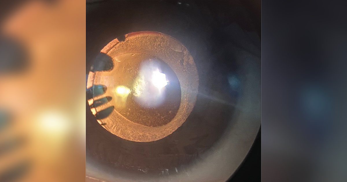
Radiation cataract is a recognized complication of radiotherapy and occupational exposure, and its importance has grown in recent years because evidence shows that the dose needed to damage the lens is lower than once thought.
What the cataract looks like
Radiation cataract starts as a small cluster of dots and vacuoles in the posterior pole of the lens, just under the capsule.
As the opacity grows, it develops into a posterior subcapsular plaque, and later it may involve the cortex.
Its appearance is similar to other forms of posterior subcapsular cataract, which is why the history of exposure is the main clue.
Symptoms are the same as for any posterior subcapsular cataract: glare, reduced vision in bright light, and difficulty reading (see posterior subcapsular cataract).
Why the lens is so vulnerable
Lens epithelial cells at the germinative zone divide throughout life, and their daughter cells migrate to the posterior pole and differentiate into fibers.
Cells that carry radiation-induced DNA damage and cannot clear it migrate there and form opaque fibers.
Because the lens does not shed cells, the damage stays and accumulates, and the opacity appears months to years after exposure.
All-fit Slit-Lamp Adapter
Record and share exactly what you see at the slit lamp. One adapter fits any slit lamp or surgical microscope — and any smartphone.
From Choroida — the team behind this siteDose and latency
The more radiation, the shorter the latency.
After high doses, opacities may appear within a year, and after lower doses, 10 years or more may pass.
The International Commission on Radiological Protection used to put the threshold for detectable opacities at around 2 Gy for an acute exposure and 5 Gy for prolonged exposure.
In 2011, it lowered the dose limit for the lens in occupational settings, accepting that the threshold may be about 0.5 Gy and that a “no threshold” model is possible.
Who is exposed
- Patients treated with radiotherapy for head and neck cancer, orbital tumors, or brain tumors, particularly if the lens was not shielded
- Patients receiving total body irradiation before stem cell transplantation, in whom cataract is frequent (see ocular graft-versus-host disease)
- Patients who have had plaque or proton beam therapy for ocular tumors (see plaque brachytherapy)
- Interventional cardiologists, radiologists, and operating theatre staff who work near an X-ray source without eye protection
- Aircrew and astronauts, exposed to cosmic radiation
- Survivors of atomic bomb exposure, among whom the relationship between dose and cataract was first documented
Prevention
Radiation protection principles apply: keep the exposure as low as reasonably achievable, use lead glasses with side shields when working in the fluoroscopy suite, use ceiling-mounted shields, and maintain distance from the source.
In radiotherapy, planning that spares the lens, where possible, helps.
Management
Observation is appropriate for early opacities.
When the cataract causes visual symptoms, cataract surgery gives results comparable to other forms of cataract.
Patients who were treated with radiation should have a periodic examination for other radiation effects, including dry eye and radiation retinopathy (see radiation keratopathy).
Counselling patients and staff
Patients who receive radiotherapy near the eye should be told that cataract is a possible late effect, and that vision changes should be reported. For staff who work with radiation, an annual eye examination is a reasonable precaution, along with the use of protective glasses and lead shielding. Employers should keep records of the doses received, and clinicians should ask about the occupational history when they see a young patient with posterior subcapsular cataract.
Surgical considerations
Cataract surgery after radiation is usually straightforward, but the surgeon should look for dry eye, which may be severe, for retinal radiation damage, and for zonular weakness in eyes that received high doses. Postoperative inflammation may be more pronounced, and healing may be slower in irradiated tissues. Explain that the visual result will depend on the state of the retina and the optic nerve, and not only on the lens.
What the lens looks like early on
The first sign may be a tiny cluster of dots and vacuoles at the posterior pole, visible only by retroillumination after dilation. Later, a granular, saucer-shaped plaque forms, which grows and may involve the cortex. Visual acuity may be normal at this stage, even though glare and difficulty in bright light are already present. Contrast sensitivity and glare testing may reveal a deficit before the Snellen chart does, and they are useful for patients with an occupational exposure.
Prognosis
Visual outcome after surgery is good, though the underlying condition and any retinal damage from radiation may limit the final result.


Document what you see
Two smartphone imaging tools built for everyday clinic use — one for the slit lamp, one for the fundus.
From Choroida — the team behind this siteReferences
- Ainsbury EA, Barnard S, Bright S, et al. Ionizing radiation induced cataracts: recent biological and mechanistic developments and perspectives for future research. Mutat Res Rev Mutat Res. 2016;770:238-261.
- ICRP. Statement on tissue reactions / early and late effects of radiation in normal tissues and organs. ICRP Publication 118. Ann ICRP. 2012;41(1/2).
- Chodick G, Bekiroglu N, Hauptmann M, et al. Risk of cataract after exposure to low doses of ionizing radiation: a 20-year prospective cohort study among US radiologic technologists. Am J Epidemiol. 2008;168:620-631.
- Worgul BV, Kundiyev YI, Sergiyenko NM, et al. Cataracts among Chernobyl clean-up workers: implications regarding permissible eye exposures. Radiat Res. 2007;167:233-243.
Test yourself
A few questions straight from this article.
-
Where in the lens does a radiation cataract first appear?
It begins as a small cluster of dots and vacuoles at the posterior pole just beneath the capsule, later becoming a posterior subcapsular plaque that may involve the cortex. -
Why does radiation damage to the lens accumulate instead of clearing?
Because the lens does not shed cells, radiation-damaged cells remain and the damage accumulates, so the opacity appears months to years after the exposure. -
Which lens cells give rise to the opaque fibres of a radiation cataract?
Lens epithelial cells at the germinative zone divide throughout life; daughter cells carrying unrepaired radiation DNA damage migrate to the posterior pole and differentiate into opaque fibres. -
In 2011 the International Commission on Radiological Protection accepted a lens threshold as low as which dose?
Having previously placed the threshold near 2 Gy for acute and 5 Gy for prolonged exposure, in 2011 the Commission lowered the occupational dose limit and accepted a threshold of about 0.5 Gy, with a no-threshold model possible. -
How does the radiation dose relate to the latency of a radiation cataract?
The more radiation received, the shorter the latency: after high doses opacities may appear within a year, while after lower doses ten years or more may pass. -
Which occupational group is at risk of radiation cataract from working near an X-ray source?
Interventional cardiologists, radiologists and operating theatre staff who work near an X-ray source without eye protection are among the exposed groups. -
What is the main clue that a posterior subcapsular cataract is radiation-induced?
Radiation cataract looks like other forms of posterior subcapsular cataract, so the history of exposure is the main clue to its cause. -
Which eye protection is recommended for staff working in a fluoroscopy suite?
Protection rests on keeping exposure as low as reasonably achievable, lead glasses with side shields, ceiling-mounted shields, and distance from the source. -
Which lens problem should be sought before cataract surgery in an eye given high-dose radiation?
Surgery is usually straightforward, but the surgeon should look for severe dry eye, retinal radiation damage and zonular weakness in eyes that received high doses. -
Which test may reveal a deficit in radiation cataract before Snellen acuity falls?
Contrast sensitivity and glare testing may show a deficit before the Snellen chart does, which makes them useful in patients with occupational exposure.
