
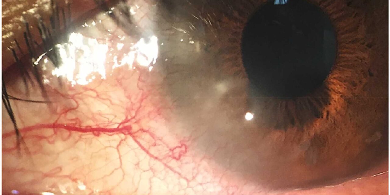
Pediatric blepharokeratoconjunctivitis is a condition that tends to be under-recognized for a specific, understandable reason: chronic eyelid and ocular surface inflammation in children is often attributed to recurrent, individually treated episodes of conjunctivitis or blepharitis, rather than being connected together as a single, chronic underlying disease process that, left inadequately managed, carries a real risk of progressive corneal scarring and vascularization over time.
A Chronic Disease Often Mistaken for a Series of Acute Ones
This condition represents chronic inflammation of the eyelid margins, meibomian glands, conjunctiva, and cornea in children, and because individual flares can resemble an ordinary, self-limited episode of blepharitis or conjunctivitis, the chronic, recurrent, underlying nature of the disease is often missed, particularly when each episode is evaluated and treated in isolation without recognizing the broader, ongoing pattern.
Clinical Features
- Chronic or recurrent lid margin inflammation, with erythema, telangiectasia, and often meibomian gland dysfunction
- Recurrent chalazia are a common and sometimes prominent presenting feature, and a child with genuinely recurrent chalazia, rather than a single isolated episode, should prompt consideration of this broader underlying chronic disease process
- Conjunctival injection and a papillary or follicular conjunctival reaction
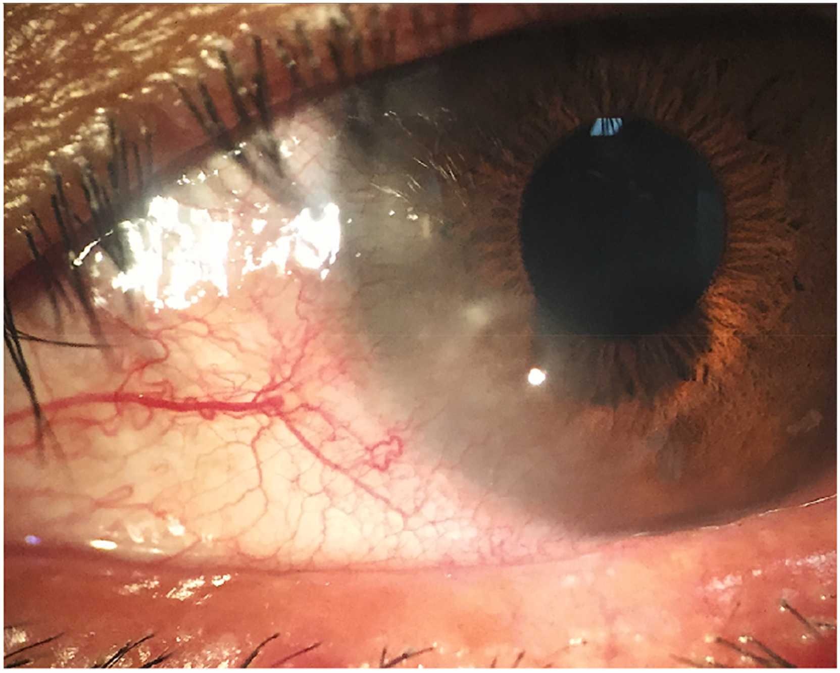
- Corneal involvement, ranging from peripheral punctate keratopathy and marginal infiltrates to, in more severe or longstanding cases, corneal vascularization and scarring, representing the most visually significant potential consequence of inadequately controlled disease
- Photophobia, tearing, and eye rubbing are common, nonspecific symptoms that can be easily attributed to other, more commonly considered pediatric eye conditions if this specific diagnosis is not considered
All-fit Slit-Lamp Adapter
Record and share exactly what you see at the slit lamp. One adapter fits any slit lamp or surgical microscope — and any smartphone.
From Choroida — the team behind this siteWhy Under-Recognition Matters
Because individual flares can be managed symptomatically without the broader chronic pattern being recognized, some children experience ongoing, low-grade disease activity over months to years, during which progressive corneal changes, including vascularization and scarring, can develop gradually and sometimes reach a visually significant degree before the underlying chronic condition is finally correctly identified and more comprehensively addressed.
Diagnostic Evaluation
- A careful history specifically asking about the frequency and pattern of prior episodes, including recurrent chalazia, recurrent “pink eye,” or recurrent eyelid inflammation, since connecting these prior episodes together is often the key step in reaching the correct underlying diagnosis
- Slit-lamp examination characterizing lid margin, meibomian gland, conjunctival, and corneal findings, with particular attention to any corneal vascularization, infiltrates, or scarring that would indicate more significant, longer-standing disease activity
- Assessment for associated skin conditions, including rosacea or seborrheic dermatitis, which can be associated with this condition, similar to the broader association seen between these skin conditions and blepharokeratoconjunctivitis in adults
Management
Lid Hygiene
Regular, consistent lid hygiene, including warm compresses and gentle lid margin cleansing, forms the foundation of management, aiming to reduce meibomian gland dysfunction and the bacterial and inflammatory burden at the lid margin that drives ongoing disease activity.
Topical and Oral Antibiotic Therapy
- Topical antibiotic or antibiotic-corticosteroid combination therapy is used for active flares and to help reduce lid margin bacterial colonization
- Oral antibiotics, particularly from the tetracycline class in older children (used with appropriate attention to age-related contraindications) or azithromycin in younger children, are sometimes used for more significant or recurrent disease, given their additional anti-inflammatory properties beyond their antimicrobial effect
Managing Corneal Involvement
Topical corticosteroids are used for active corneal inflammation or marginal infiltrates, with careful monitoring given the particular considerations relevant to pediatric corticosteroid use, including intraocular pressure monitoring.
Long-Term, Sustained Management
Because this is a chronic condition with a tendency toward recurrence, sustained, ongoing lid hygiene and periodic reassessment, rather than treatment limited to acute flares alone, is central to preventing the cumulative corneal complications that inadequately controlled, longstanding disease activity can produce.
What to teach the family
Show the parents how to apply a warm compress for several minutes and how to massage and clean the lids gently, using a clean cloth or lid wipes. Explain that this is a daily routine, like brushing teeth, and that it must be continued for months. Give written instructions, and describe the signs of a flare, such as redness, sticky lids, or light sensitivity.
When to refer
Refer children with corneal vascularization, scarring, or poor vision, since amblyopia may follow if the cornea is involved. Any child with a recurrent chalazion should be examined for this condition. Take photographs of the lids and cornea to monitor the response.
Prognosis
With early recognition, connecting a pattern of recurrent episodes into the correct chronic diagnosis, and consistent, sustained management, most children with blepharokeratoconjunctivitis avoid significant corneal complications and maintain good long-term vision.
Delayed recognition, allowing ongoing, inadequately controlled disease activity to persist over an extended period, carries a real risk of progressive corneal vascularization and scarring, underscoring the value of considering this diagnosis specifically in any child with a pattern of recurrent lid, conjunctival, or corneal inflammatory episodes rather than treating each occurrence as an isolated, unrelated event.


Document what you see
Two smartphone imaging tools built for everyday clinic use — one for the slit lamp, one for the fundus.
From Choroida — the team behind this siteReferences
- Jones SM, Weinstein JM, Cumberland P, Klein N, Nischal KK. Visual outcome and corneal changes in children with chronic blepharokeratoconjunctivitis. Ophthalmology. 2007;114:2271-2280.
- Viswalingam M, Rauz S, Morlet N, Dart JK. Blepharokeratoconjunctivitis in children: diagnosis and treatment. Br J Ophthalmol. 2005;89:400-403.
- Doan S. Blepharokeratoconjunctivitis in children. J Fr Ophtalmol. 2014;37:642-647.
- Gokhale NS. Blepharokeratoconjunctivitis in children. J AAPOS. 2014;18:317-318.
Test yourself
A few questions straight from this article.
-
Which structures are chronically inflamed in pediatric blepharokeratoconjunctivitis?
The condition is chronic inflammation of the eyelid margins, meibomian glands, conjunctiva and cornea in children, rather than a series of unrelated acute events. -
Why is pediatric blepharokeratoconjunctivitis so often under-recognised?
Because each flare can look like a self-limited episode of blepharitis or conjunctivitis, the chronic underlying pattern is missed when episodes are treated in isolation. -
Which recurrent eyelid lesion should prompt consideration of pediatric blepharokeratoconjunctivitis?
Recurrent chalazia are a common and sometimes prominent presenting feature, and a child with genuinely recurrent chalazia should prompt consideration of this chronic disease. -
Which corneal change is the most visually significant consequence of poorly controlled disease?
Corneal involvement can progress from punctate keratopathy and marginal infiltrates to vascularization and scarring, the most visually significant consequence of inadequately controlled disease. -
Which conjunctival reaction is typically found in pediatric blepharokeratoconjunctivitis?
Conjunctival injection with a papillary or follicular conjunctival reaction is among the characteristic clinical features. -
Which associated skin conditions should be assessed for in an affected child?
Assessment includes looking for associated skin conditions such as rosacea or seborrhoeic dermatitis, which can accompany this condition. -
What forms the foundation of management in pediatric blepharokeratoconjunctivitis?
Consistent lid hygiene with warm compresses and gentle lid margin cleansing is the foundation of management, reducing meibomian gland dysfunction and lid margin inflammatory burden. -
Which oral antibiotic is used in younger children with significant or recurrent disease?
Tetracycline-class drugs are used in older children with attention to age-related contraindications, while azithromycin is used in younger children. -
When topical corticosteroids treat corneal inflammation in a child, what monitoring is required?
Topical corticosteroids for active corneal inflammation or marginal infiltrates need careful monitoring, including intraocular pressure monitoring, in paediatric use. -
What most determines whether an affected child avoids significant corneal complications?
Most children avoid significant corneal complications and keep good long-term vision when recurrent episodes are recognised as one chronic diagnosis and managed consistently.
