
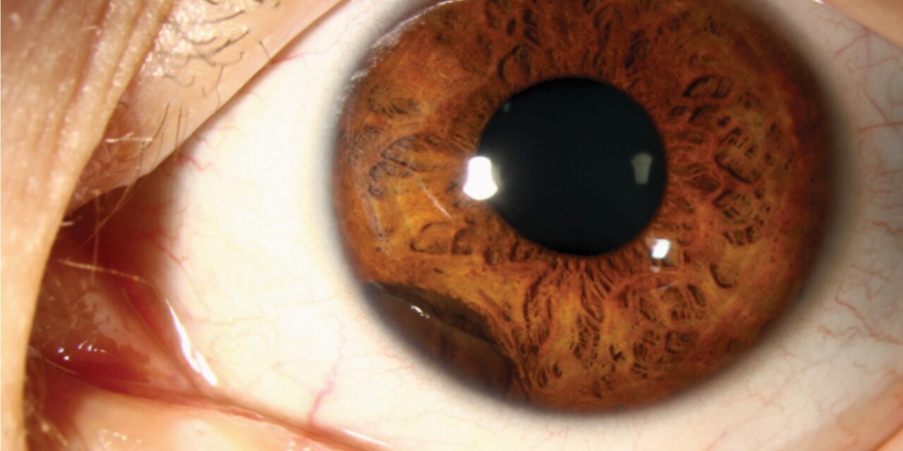
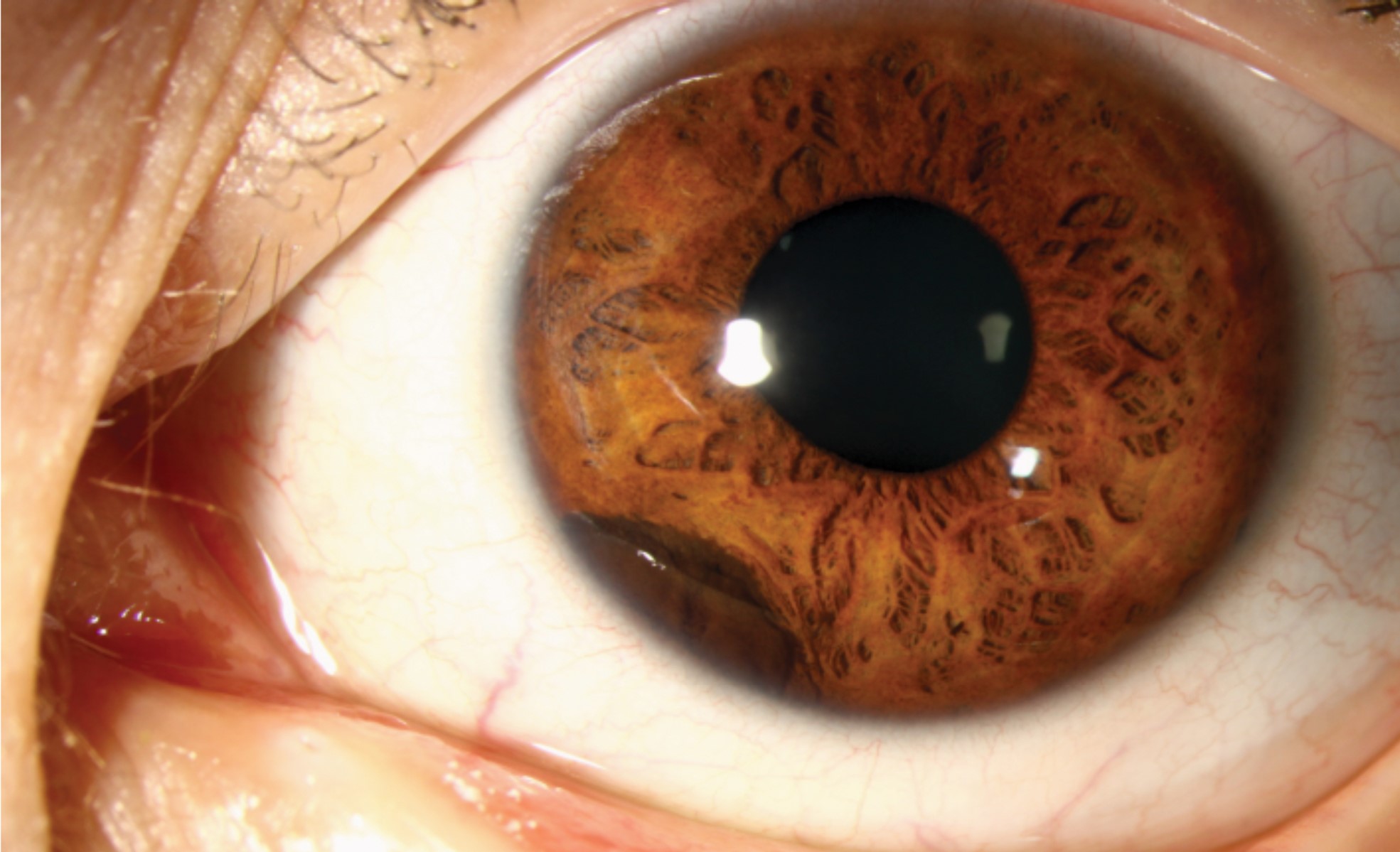
A ciliary body cyst sits in a genuinely hard-to-reach location, directly behind the iris, which is precisely why these lesions are so often entirely invisible on standard slit-lamp examination and are instead discovered incidentally, either through dilated examination revealing a subtle iris bulge, or, more definitively, through ultrasound biomicroscopy, a specialized imaging technique capable of seeing behind the iris to where routine examination simply cannot reach.
Why These Cysts Are So Often Missed
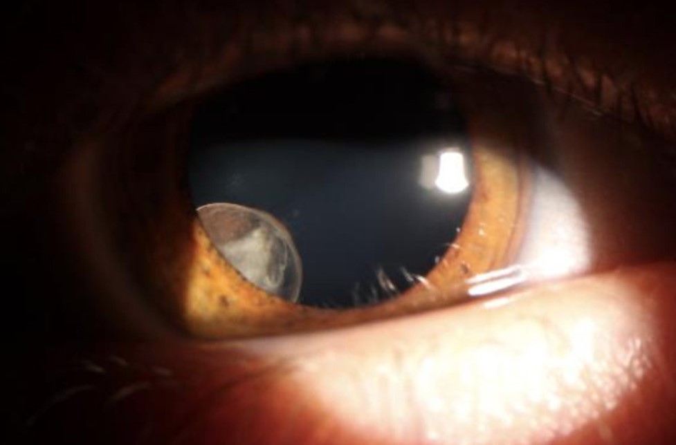
The ciliary body, and specifically the pars plicata portion where these cysts most commonly arise, sits anatomically behind the iris, outside the field of view of a standard slit-lamp examination, even with pupillary dilation, unless the cyst is large enough to cause a visible secondary change, such as localized iris bulging or angle narrowing, that draws attention to the underlying, otherwise hidden lesion.
Types of Ciliary Body Cysts
Primary (Neuroepithelial) Cysts
These arise from the neuroepithelial layers of the ciliary body itself, are generally congenital or develop without an identifiable external cause, and are typically benign, stable, and asymptomatic throughout a patient’s life.
Secondary Cysts
These can arise in association with certain medications (similar to the mechanism described for some iris cysts), prior trauma, or chronic intraocular inflammation, reflecting a different, acquired underlying process rather than a primary developmental origin (see iris cyst classification for the related discussion of similar mechanisms producing cysts at the iris itself).
All-fit Slit-Lamp Adapter
Record and share exactly what you see at the slit lamp. One adapter fits any slit lamp or surgical microscope — and any smartphone.
From Choroida — the team behind this siteClinical Presentation
- The great majority of ciliary body cysts are entirely asymptomatic and are discovered incidentally, either through indirect signs on examination or through imaging performed for another reason
- Larger cysts can occasionally cause a visible, localized bulging of the peripheral iris, sometimes noted on careful slit-lamp examination even without direct visualization of the cyst itself
- In uncommon cases, larger or multiple cysts can contribute to secondary angle narrowing or, rarely, angle closure, by mechanically crowding the anterior chamber angle from behind
- Refractive changes, including induced astigmatism, have been described in rare cases where a cyst is large enough to mechanically distort adjacent structures
Diagnostic Evaluation
- Ultrasound biomicroscopy is the key imaging tool for directly visualizing ciliary body cysts, given their location behind the iris, and is considered the gold standard for confirming and characterizing this diagnosis
- Gonioscopy can sometimes reveal indirect signs, including localized angle narrowing or iris bulging, that prompt further investigation for an underlying ciliary body cyst
- Transillumination can occasionally support the diagnosis when a cyst is large enough and appropriately positioned, though it is a less reliable tool for this specific, more posteriorly located lesion compared with its usefulness for iris lesions
Distinguishing From a Solid Ciliary Body Tumor
As with iris cysts, distinguishing a ciliary body cyst from a solid ciliary body tumor, including ciliary body melanoma, is an essential diagnostic task given the very different management implications (see ciliary body melanoma for the related diagnostic approach to a solid ciliary body mass).
- Ultrasound biomicroscopy directly distinguishes the smooth-walled, echolucent (fluid-filled) appearance of a cyst from the solid, echogenic internal pattern of a tumor
- The generally stable, non-growing natural history of most ciliary body cysts, confirmed on serial examination when needed, further supports a benign cystic diagnosis over a concerning solid lesion
Management
Observation
The great majority of ciliary body cysts, being asymptomatic and stable, require no treatment and are managed with simple observation, often without even the need for serial monitoring once the benign nature of the finding is confirmed.
Intervention for Symptomatic or Complicated Cysts
For the uncommon cases where a cyst is large enough to cause secondary angle narrowing, contribute to elevated intraocular pressure, or cause other symptomatic mechanical effects, treatment options include laser therapy or, less commonly, surgical drainage or excision, reserved specifically for cysts causing a genuine clinical problem rather than applied routinely to an incidentally discovered, asymptomatic lesion.
Practical follow-up
Most ciliary body cysts are found incidentally, are stable, and need no treatment. Record the size and position with ultrasound biomicroscopy, and review at an interval if the lesion is large, since growth would suggest a different diagnosis.
Prognosis
Ciliary body cysts, in the great majority of cases, have an excellent prognosis and remain entirely stable and asymptomatic throughout a patient’s life, requiring nothing beyond the reassurance that comes with a confirmed benign diagnosis.
The main clinical value in correctly identifying and characterizing a ciliary body cyst lies in confidently distinguishing it from a solid tumor requiring active monitoring or intervention, which is exactly the distinction ultrasound biomicroscopy is so well suited to making.


Document what you see
Two smartphone imaging tools built for everyday clinic use — one for the slit lamp, one for the fundus.
From Choroida — the team behind this siteReferences
- Marigo FA, Finger PT, McCormick SA, Iacob CE, Grossniklaus HE, Bekerman VP. Iris and ciliary body melanomas: ultrasound biomicroscopy for the diagnosis and management. Ophthalmology. 2000;107:1929-1936.
- Kunimatsu S, Araie M, Ohara K, Hamada C. Ultrasound biomicroscopy of ciliary body cysts. Am J Ophthalmol. 1999;127:48-55.
- Shields JA, Shields CL, Kiratli H, De Potter P. Peripheral cystoid degeneration of the retina. A cause of severe visual field defect. Retina. 1994;14:493-497.
- Finger PT, Perry HD, Packer S, Erdey RA, Weisman GD, Sibony PA. Pigmented and nonpigmented benign tumors of the ciliary body epithelium. Ophthalmology. 1989;96:1721-1726.
Test yourself
A few questions straight from this article.
-
Ciliary body cysts most commonly arise from which part of the ciliary body?
These cysts arise most commonly from the pars plicata portion of the ciliary body, which lies anatomically behind the iris. -
Why does standard slit-lamp examination usually fail to show a ciliary body cyst?
The cyst sits behind the iris, outside the field of a standard slit-lamp examination even with pupillary dilation, unless it causes a visible secondary change. -
Which imaging modality is the gold standard for confirming a ciliary body cyst?
Ultrasound biomicroscopy can see behind the iris and is considered the gold standard for confirming and characterising a ciliary body cyst. -
Primary ciliary body cysts arise from which tissue?
Primary cysts arise from the neuroepithelial layers of the ciliary body itself and are generally congenital or develop without an identifiable external cause. -
Secondary ciliary body cysts are associated with which set of factors?
Secondary cysts reflect an acquired process and can arise in association with certain medications, prior trauma, or chronic intraocular inflammation. -
How can large or multiple ciliary body cysts contribute to angle closure?
In uncommon cases larger or multiple cysts crowd the anterior chamber angle mechanically from behind, producing secondary angle narrowing or, rarely, angle closure. -
Which ultrasound biomicroscopy appearance separates a ciliary body cyst from a solid tumour?
Ultrasound biomicroscopy shows the smooth-walled, echolucent, fluid-filled appearance of a cyst, in contrast to the solid, echogenic internal pattern of a tumour. -
What is the prognosis of a confirmed ciliary body cyst in the great majority of cases?
In the great majority of cases these cysts remain entirely stable and asymptomatic throughout life, needing nothing beyond reassurance once the benign diagnosis is confirmed. -
How is a ciliary body cyst causing secondary angle narrowing and raised pressure treated?
Treatment is reserved for cysts causing a genuine clinical problem, and options then include laser therapy or, less commonly, surgical drainage or excision. -
What would documented growth of a presumed ciliary body cyst suggest?
Most ciliary body cysts are stable and non-growing, so growth on serial ultrasound biomicroscopy would point away from a benign cyst towards a different diagnosis.
