
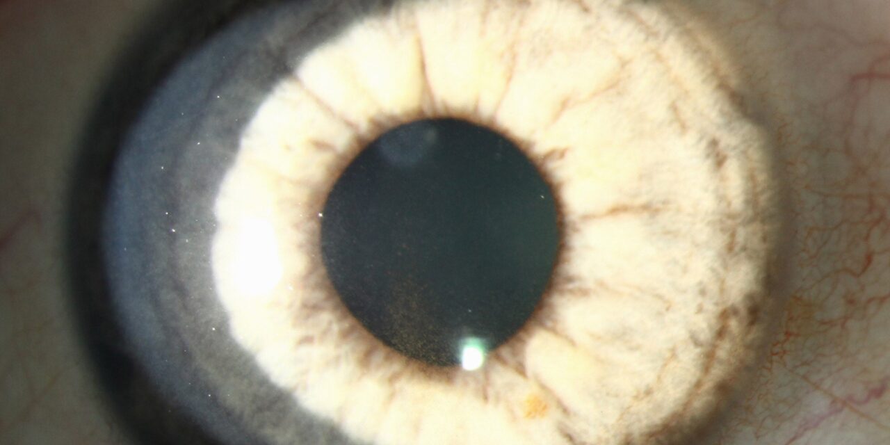
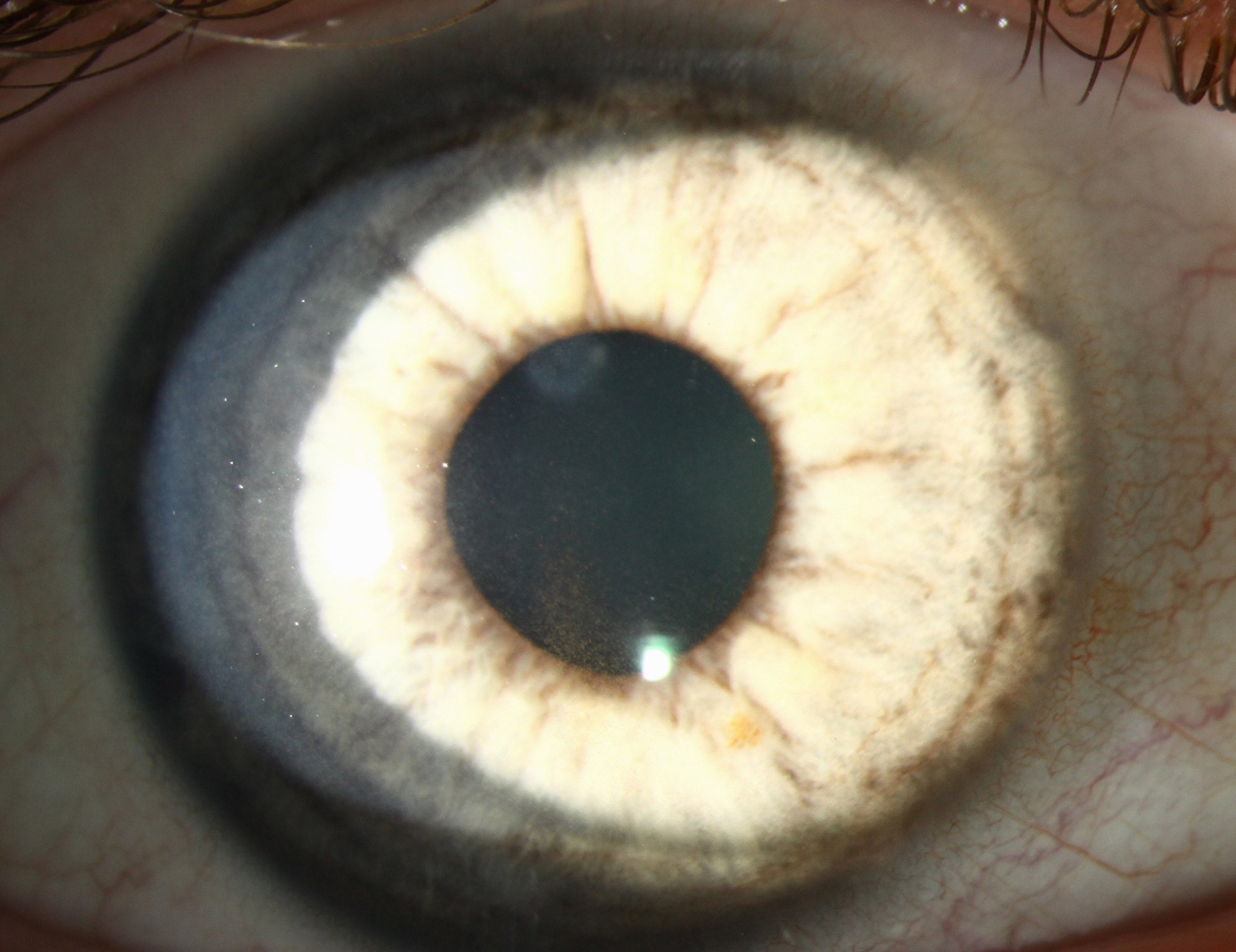
Pigmentary glaucoma is the point at which pigment dispersion syndrome, previously just an anatomic finding, becomes a genuine threat to the optic nerve, and understanding what tips a given patient from the syndrome into actual glaucoma, and recognizing the distinctive pattern of pressure spikes this condition can produce, shapes both how these patients are monitored and how urgently treatment is pursued.
From Syndrome to Disease
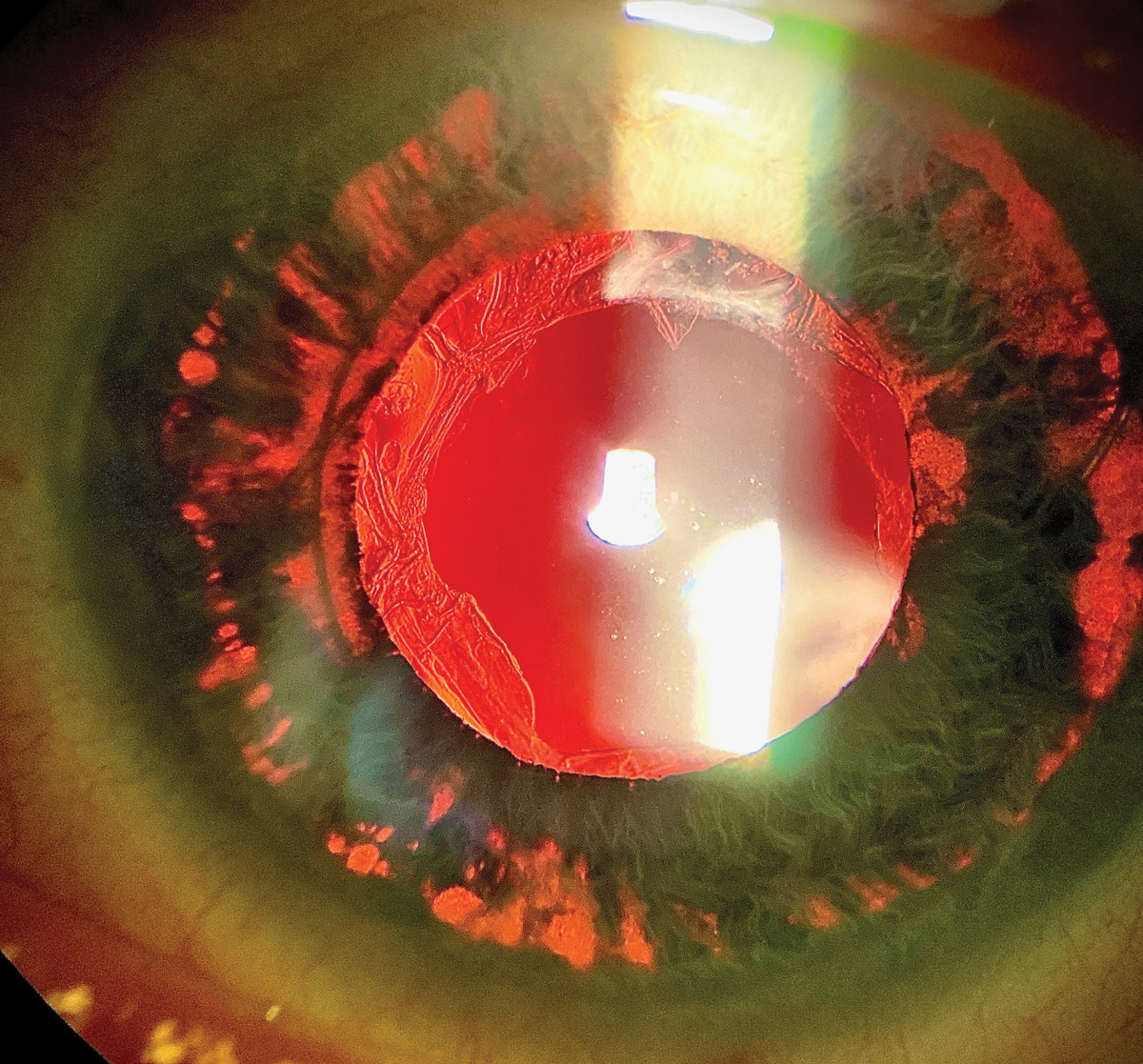
Pigment dispersion syndrome describes the anatomic triad of iris transillumination defects, Krukenberg spindle, and increased trabecular meshwork pigmentation, resulting from chronic mechanical rubbing between the posterior iris and anterior zonular fibers (see Krukenberg spindle and trabecular meshwork pigmentation for the individual components of this triad).
Pigmentary glaucoma develops when the pigment deposited within the trabecular meshwork, over time, begins to impair normal aqueous outflow enough to elevate intraocular pressure and, eventually, cause optic nerve damage, a progression that occurs in a meaningful but not universal proportion of patients with the underlying syndrome.
Who Develops Pigmentary Glaucoma
- Young, myopic patients are disproportionately affected, and this demographic pattern, notably different from the older, more hyperopic-leaning population typical of primary open-angle glaucoma, is itself a useful clinical clue
- Men are affected more frequently than women in most reported series
- Not every patient with pigment dispersion syndrome develops glaucoma, and the specific factors determining which patients progress from syndrome to disease are not fully understood, supporting the value of ongoing monitoring in every patient with the underlying syndrome rather than assuming a uniform, predictable course
All-fit Slit-Lamp Adapter
Record and share exactly what you see at the slit lamp. One adapter fits any slit lamp or surgical microscope — and any smartphone.
From Choroida — the team behind this siteThe Distinctive Exercise-Related Pressure Spike
A recognized and clinically important feature of pigmentary glaucoma is the tendency for intraocular pressure to spike acutely following physical exercise, particularly exercise involving rapid or repetitive eye movement, such as running or jumping, thought to relate to mechanical iris movement during exercise liberating an additional surge of pigment into the aqueous outflow pathway.
- This pattern means that a single, routine in-office pressure measurement, taken under resting conditions, can substantially underestimate a patient’s true peak pressure burden over the course of a day that includes physical activity
- Asking specifically about exercise habits, and considering pressure checks at different times or after exertion in select patients, can be a valuable part of fully characterizing disease severity in this specific population
Clinical Features
- Elevated intraocular pressure, sometimes with documented spikes well above what a single resting office measurement suggests
- Optic nerve and visual field changes consistent with glaucomatous damage, assessed using the same standard techniques applied to other forms of open-angle glaucoma
- The characteristic pigment dispersion triad findings (Krukenberg spindle, iris transillumination defects, trabecular pigmentation) remain present and are part of confirming the underlying syndrome driving the secondary glaucoma
- As patients age, pigment liberation and the associated clinical findings sometimes lessen somewhat, related to age-related changes in iris configuration, though established glaucomatous optic nerve damage does not reverse even if the underlying pigment dispersion activity itself later diminishes
Diagnostic Evaluation
- Gonioscopy and slit-lamp examination confirming the underlying pigment dispersion syndrome findings
- Intraocular pressure monitoring, with consideration of the exercise-related spike pattern when characterizing a given patient’s true pressure burden
- Optic nerve assessment and visual field testing, following standard glaucoma evaluation principles, to determine whether structural or functional glaucomatous damage has developed
- Refractive assessment, given the recognized association with myopia in this specific glaucoma subtype
Management
Medical Therapy
Topical aqueous-suppressant medications are generally used as first-line treatment, similar to management of primary open-angle glaucoma, aiming to reduce intraocular pressure and protect the optic nerve.
- Miotic agents, including pilocarpine, have a specific rationale in pigmentary glaucoma beyond their general pressure-lowering effect, since they also cause pupillary constriction that reduces the mechanical iris-zonular contact believed to drive ongoing pigment release, directly addressing the underlying mechanism rather than only the resulting pressure elevation
Laser Peripheral Iridotomy
Creating a peripheral iridotomy can help flatten the iris and reduce the posterior bowing that contributes to iris-zonular contact, theoretically reducing ongoing pigment liberation, and is used in appropriately selected patients, though its role and effectiveness specifically for altering the long-term course of pigmentary glaucoma remains an area of ongoing clinical judgment and discussion.
Laser Trabeculoplasty and Surgery
Selective or argon laser trabeculoplasty can be effective for pressure reduction in pigmentary glaucoma, and incisional glaucoma surgery, including trabeculectomy or drainage device implantation, is reserved for disease not adequately controlled with medical and laser therapy, following standard escalation principles used across open-angle glaucoma management more broadly.
Prognosis
With appropriate, sustained management, including attention to the distinctive exercise-related pressure spike pattern and its implications for true disease severity assessment, many patients with pigmentary glaucoma achieve good long-term pressure control and stable visual function.
Given the relatively young age at which this condition often affects patients, a long treatment horizon and sustained, engaged monitoring are particularly important considerations in counseling and managing this specific glaucoma population.


Document what you see
Two smartphone imaging tools built for everyday clinic use — one for the slit lamp, one for the fundus.
From Choroida — the team behind this siteReferences
- Farrar SM, Shields MB. Current concepts in pigmentary glaucoma. Surv Ophthalmol. 1993;37:233-252.
- Haynes WL, Johnson AT, Alward WL. Effects of jogging exercise on patients with the pigmentary dispersion syndrome and pigmentary glaucoma. Ophthalmology. 1992;99:1096-1103.
- Richter CU, Richardson TM, Grant WM. Pigmentary dispersion syndrome and pigmentary glaucoma. A prospective study of the natural history. Arch Ophthalmol. 1986;104:211-215.
- Niyadurupola N, Broadway DC. Pigment dispersion syndrome and pigmentary glaucoma–a major review. Clin Exp Ophthalmol. 2008;36:868-882.
Test yourself
A few questions straight from this article.
-
Pigment dispersion syndrome is said to have become pigmentary glaucoma when which change occurs?
The syndrome is an anatomic finding only; glaucoma is diagnosed once pigment in the trabecular meshwork impairs aqueous outflow enough to raise pressure and eventually injure the optic nerve. -
Which three findings make up the anatomic triad of pigment dispersion syndrome?
Pigment dispersion syndrome is defined by iris transillumination defects, a Krukenberg spindle, and increased trabecular meshwork pigmentation. -
Pigment release in pigment dispersion syndrome results from chronic rubbing between which two structures?
The triad arises from chronic mechanical rubbing of the posterior iris against the anterior zonular fibres, which liberates pigment into the aqueous. -
Which patient profile is disproportionately affected by pigmentary glaucoma?
Pigmentary glaucoma favours young myopic patients and men, a pattern notably different from the older, more hyperopic-leaning population typical of primary open-angle glaucoma. -
In pigmentary glaucoma, intraocular pressure characteristically spikes acutely after what?
Running or jumping moves the iris mechanically and liberates a surge of pigment into the outflow pathway, producing an acute post-exercise pressure spike. -
What is the main practical consequence of the exercise-related pressure spike in pigmentary glaucoma?
Because the spike follows exertion, one routine resting measurement can substantially understate the patient's true peak pressure burden across an active day. -
Why do miotic agents such as pilocarpine have a specific rationale in pigmentary glaucoma?
Beyond lowering pressure, miotics constrict the pupil and cut down the mechanical iris-zonular contact that drives ongoing pigment release, addressing the mechanism itself. -
How is laser peripheral iridotomy thought to help in pigmentary glaucoma?
A peripheral iridotomy flattens the iris and reduces the posterior bowing that brings it into contact with the zonules, theoretically limiting further pigment liberation. -
What happens to pigmentary glaucoma findings as affected patients grow older?
Age-related changes in iris configuration can reduce pigment liberation and its clinical signs, yet glaucomatous optic nerve damage already established does not reverse. -
What is generally used as first-line treatment for raised pressure in pigmentary glaucoma?
As in primary open-angle glaucoma, topical aqueous suppressants are first-line, with laser and then incisional surgery reserved for inadequately controlled disease.
