
Contact lens-induced acute red eye, abbreviated CLARE, produces a presentation dramatic enough to worry both patient and clinician, sudden, severe pain and redness typically noticed on waking, and the single most important thing to get right about this condition is recognizing it as sterile inflammation rather than infection, since treating it as though it were microbial keratitis leads to unnecessary, more aggressive, and ultimately unhelpful treatment.
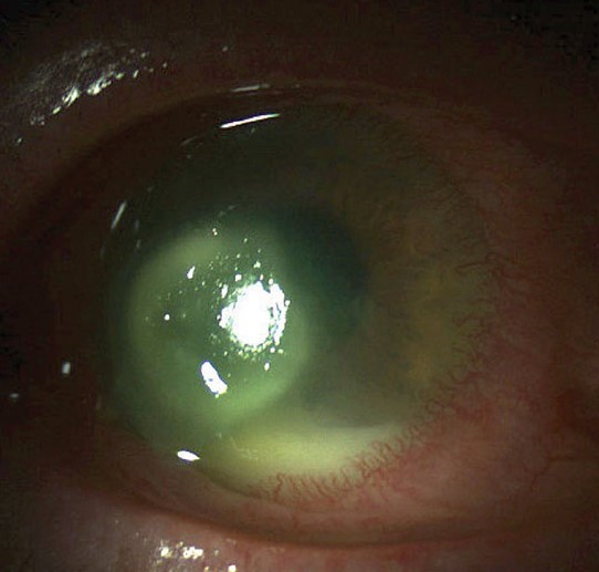
What’s Actually Happening
CLARE is a sterile inflammatory reaction believed to result from an immune response to bacterial byproducts, particularly endotoxins from gram-negative organisms such as certain strains that can colonize contact lenses and the space between the lens and cornea, rather than from direct bacterial invasion of the cornea itself.
This distinction, an inflammatory reaction to bacterial products rather than active bacterial infection of corneal tissue, is exactly why CLARE is fundamentally a different disease process from infectious contact lens-related keratitis, despite both occurring in contact lens wearers and both producing an acutely painful red eye.
Risk Factors
- Overnight or extended contact lens wear is the dominant risk factor, since prolonged, uninterrupted lens wear allows the bacterial colonization believed to drive this reaction to build up to a greater degree
- A pattern of recurrent episodes in the same patient, sometimes related to a particular lens type, wearing schedule, or lens care routine
All-fit Slit-Lamp Adapter
Record and share exactly what you see at the slit lamp. One adapter fits any slit lamp or surgical microscope — and any smartphone.
From Choroida — the team behind this siteClinical Presentation
- Sudden onset of significant eye pain, redness, and photophobia, classically noticed upon waking after a period of overnight lens wear
- One or more peripheral or mid-peripheral corneal infiltrates, generally smaller and more superficial than the dense, often larger infiltrate typical of infectious microbial keratitis
- A notable absence of significant epithelial defect overlying the infiltrate in many cases, in contrast to the epithelial breakdown typically seen with an infectious corneal ulcer
- Symptoms and findings often improve relatively quickly, within days, once the contact lens is discontinued, a course distinctly faster than the days-to-weeks typical of an infectious process requiring antimicrobial treatment
Distinguishing CLARE From Infectious Keratitis
This distinction carries real clinical weight, since it determines whether a patient needs aggressive antimicrobial treatment or simply lens discontinuation and supportive care.
- CLARE infiltrates tend to be smaller, more peripheral, and often multiple, while infectious keratitis more typically produces a single, larger, more central or progressively enlarging infiltrate
- The characteristic absence of a significant overlying epithelial defect in CLARE contrasts with the epithelial breakdown generally expected over an infectious infiltrate
- CLARE typically shows rapid improvement within a few days of lens discontinuation, while infectious keratitis would be expected to continue progressing without appropriate antimicrobial treatment
- When the distinction remains genuinely uncertain, particularly with a larger or more centrally located infiltrate, treating empirically as though infectious (with corneal culture and antibiotic coverage) until clear improvement is observed is the safer default approach, since missing a true infection carries much greater risk than briefly over-treating CLARE
Management
Lens Discontinuation
Discontinuing contact lens wear is the central, essential step in managing CLARE, since the reaction is directly tied to the lens-related colonization driving it, and improvement is generally prompt once this trigger is removed.
Supportive Care
Lubrication and, for significant discomfort, topical corticosteroids (once an infectious process has been adequately excluded or is being concurrently covered empirically) support symptomatic relief during the resolution period.
Preventing Recurrence
- Transitioning away from overnight or extended wear to daily wear with removal each night substantially reduces recurrence risk
- Reviewing and reinforcing proper lens hygiene and replacement schedule
- For patients who experience recurrent CLARE episodes despite reasonable wear modification, further evaluation of lens type, fit, and care routine is appropriate to identify and address the specific ongoing contributing factor
Why it is different from an infection
CLARE tends to appear on waking, with sudden redness, pain, and tearing, usually with small peripheral infiltrates and a quiet center. The epithelium is usually intact or has small defects. In contrast, a microbial ulcer shows a larger central or paracentral infiltrate with an epithelial defect and heavy discharge. Because the two can be hard to separate, lens wearers with infiltrates should be reviewed in a day or two, and cultured if there is any doubt.
Preventing recurrence
Stop overnight wear, replace lenses and cases regularly, avoid water exposure, and wash hands. Patients with repeated episodes should consider daily disposable lenses.
Prognosis
CLARE generally has an excellent prognosis, with prompt, often significant improvement within days of contact lens discontinuation and supportive care, and typically no lasting corneal scarring or visual impact once the episode resolves.
Recurrence is common in patients who resume the same overnight or extended wear pattern that precipitated the initial episode, which is why counseling about wear modification, not just treatment of the acute episode, is an essential part of long-term management for this specific, contact-lens-wear-driven condition.


Document what you see
Two smartphone imaging tools built for everyday clinic use — one for the slit lamp, one for the fundus.
From Choroida — the team behind this siteReferences
- Sweeney DF, Jalbert I, Covey M, et al. Clinical characterization of corneal infiltrative events observed with soft contact lens wear. Cornea. 2003;22:435-442.
- Stapleton F, Keay L, Jalbert I, Cole N. The epidemiology of contact lens related infiltrates. Optom Vis Sci. 2007;84:257-272.
- Sankaridurg PR, Willcox MD, Sharma S, et al. Haemophilus influenzae adherent to contact lenses associated with production of acute ocular inflammation. J Clin Microbiol. 1996;34:2426-2431.
- Efron N. Contact Lens Complications. 4th ed. Edinburgh: Elsevier; 2019.
Test yourself
A few questions straight from this article.
-
What process underlies contact lens-induced acute red eye (CLARE)?
CLARE is a sterile inflammatory reaction to bacterial byproducts, particularly endotoxins from gram-negative organisms colonising the lens and the space between lens and cornea, not bacterial invasion of corneal tissue. -
Which lens-wearing practice is the dominant risk factor for CLARE?
Prolonged, uninterrupted lens wear allows the bacterial colonisation believed to drive the reaction to build up to a greater degree, making overnight or extended wear the dominant risk factor. -
When does CLARE classically present?
The classic presentation is sudden significant pain, redness and photophobia noticed on waking, following a period of overnight lens wear. -
Which corneal infiltrate pattern is typical of CLARE?
CLARE produces one or more peripheral or mid-peripheral infiltrates that are generally smaller and more superficial than the dense, larger infiltrate of infectious microbial keratitis. -
Which epithelial finding helps separate CLARE from an infectious corneal ulcer?
In many CLARE cases the epithelium overlying the infiltrate is intact, contrasting with the epithelial breakdown generally expected over an infectious corneal infiltrate. -
How does the course of CLARE differ from infectious keratitis after the lens is removed?
CLARE typically shows rapid improvement within a few days of lens discontinuation, whereas infectious keratitis would be expected to continue progressing without appropriate antimicrobial treatment. -
A lens wearer has a larger, more central infiltrate and the diagnosis is genuinely uncertain. What is the safer default?
Missing a true infection carries much greater risk than briefly over-treating CLARE, so corneal culture with antibiotic coverage until clear improvement is the safer default when the distinction is uncertain. -
What is the central, essential step in managing an episode of CLARE?
Because the reaction is directly tied to lens-related colonisation, stopping lens wear is the essential step, and improvement is generally prompt once this trigger is removed. -
Which change in wearing habits most reduces the risk of recurrent CLARE?
Moving away from overnight or extended wear to daily wear with nightly removal substantially reduces recurrence, which is common in patients who resume the pattern that precipitated the episode. -
What corneal outcome is typical once an episode of CLARE resolves?
CLARE generally carries an excellent prognosis, with significant improvement within days of lens discontinuation and supportive care and typically no lasting scarring or visual consequence.
