
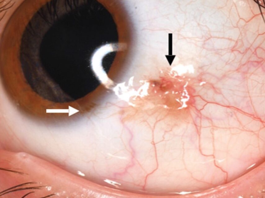
Conjunctival nevus versus melanoma requires its own distinct evaluation framework, since although both are pigmented lesions of the ocular surface, the specific features that separate a benign conjunctival nevus from a concerning melanoma or its precursor lesion, primary acquired melanosis, differ meaningfully from the risk features used to evaluate a pigmented lesion of the iris.
Conjunctival Nevus
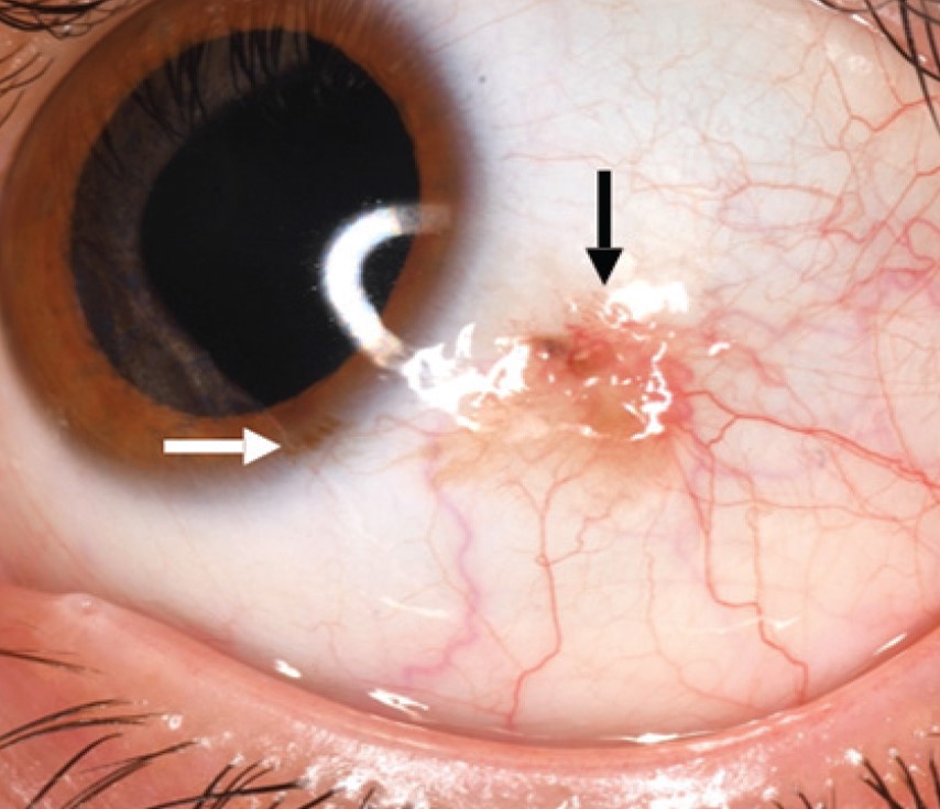
A conjunctival nevus is a common, generally benign, pigmented lesion arising within the conjunctival epithelium and substantia propria, most often located near the limbus, and often first noticed or becoming more pigmented around puberty, when hormonal changes can increase melanocyte activity within a pre-existing, previously less visible nevus.
- A highly characteristic and reassuring feature of many conjunctival nevi is the presence of small intralesional cysts, visible as clear, round inclusions within the pigmented lesion on slit-lamp examination, a finding essentially specific to benign nevi and rarely, if ever, seen within a melanoma
- Nevi are mobile over the underlying sclera, since they arise within the conjunctiva itself, which moves freely, distinguishing them from an episcleral lesion, which remains fixed beneath the mobile conjunctiva (see episcleral nevus for that related but anatomically distinct entity)
- Pigmentation can vary considerably, and some conjunctival nevi are minimally or non-pigmented (amelanotic), which can occasionally make them harder to distinguish from other conjunctival lesions on color alone
Primary Acquired Melanosis
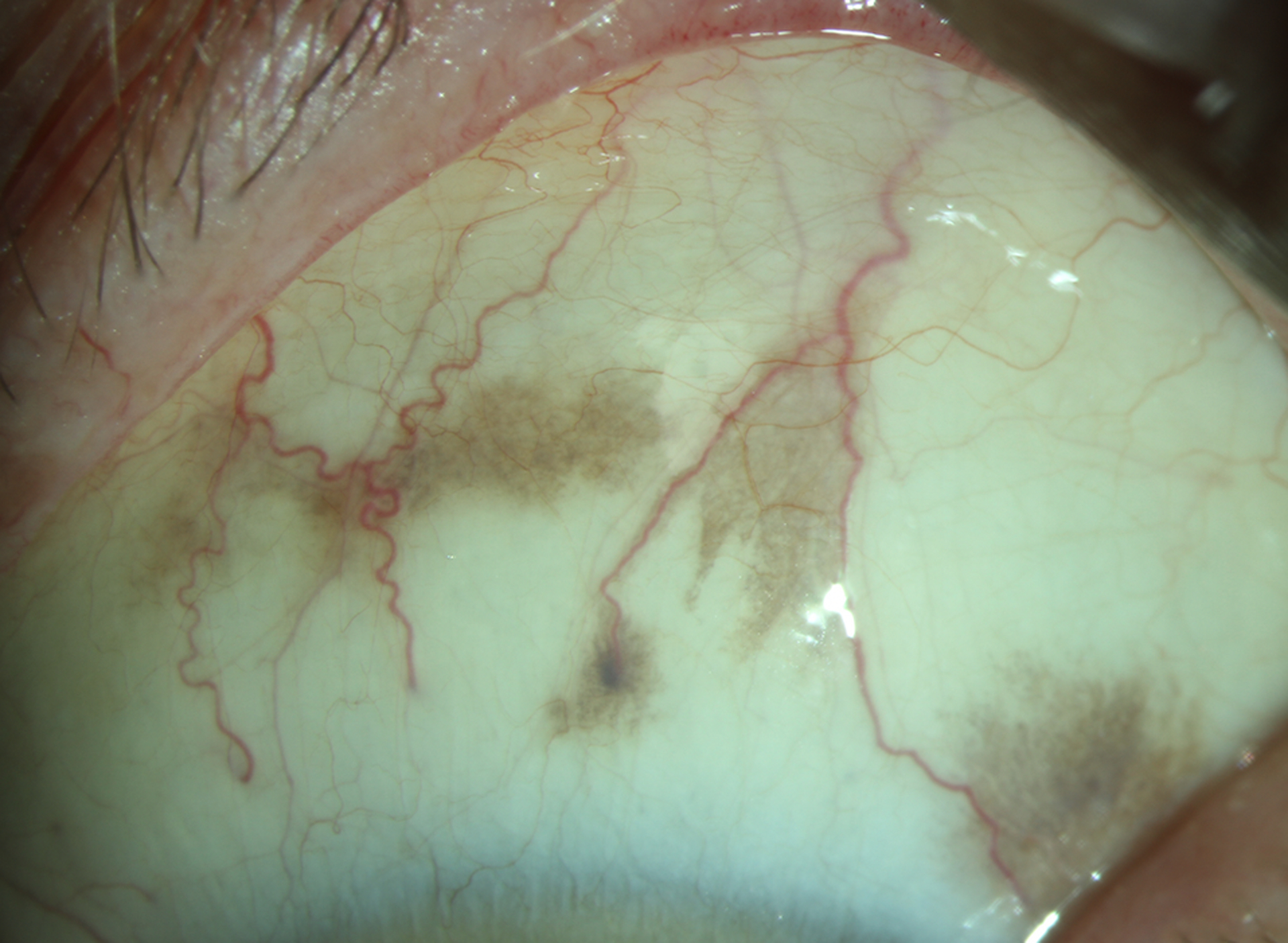
This is a distinct entity from a nevus, presenting as flat, often patchy or geographic, pigmentation of the conjunctival epithelium, typically arising later in life, and it carries genuine importance because a subset of cases, particularly those showing significant cellular atypia on biopsy, carry a meaningful risk of progression to conjunctival melanoma, making this a lesion category requiring more careful risk stratification than a typical congenital or juvenile-onset nevus.
All-fit Slit-Lamp Adapter
Record and share exactly what you see at the slit lamp. One adapter fits any slit lamp or surgical microscope — and any smartphone.
From Choroida — the team behind this siteConjunctival Melanoma
Conjunctival melanoma can arise from primary acquired melanosis with atypia, from a pre-existing nevus, or, less commonly, arise entirely de novo without a clearly identifiable precursor lesion.
- Growth, documented over serial examinations, is one of the most important risk indicators
- Prominent feeder vessels and a nodular, elevated growth pattern raise concern
- Lesions extending onto the palpebral conjunctiva, into the fornix, or onto the caruncle carry a somewhat less favorable prognosis than those confined to the more readily monitored and treated bulbar conjunctiva
- Unlike a typical nevus, melanoma and atypical primary acquired melanosis generally lack the characteristic intralesional cysts that support a benign diagnosis
Clinical Evaluation
- Careful slit-lamp examination specifically assessing for intralesional cysts, lesion mobility over the sclera, extent (including checking the palpebral conjunctiva, fornices, and caruncle, sites sometimes overlooked on a cursory examination), and any feeder vessels
- Baseline photographic documentation at first identification, supporting objective assessment of any future growth, which is often the single most important factor prompting further evaluation or intervention
- Periodic follow-up examination for lesions without immediately concerning features, with the interval determined by the initial level of suspicion
When to Biopsy
- Any lesion with documented growth, new feeder vessels, loss of mobility (suggesting invasion into deeper tissue), or other atypical features on serial examination warrants biopsy
- Primary acquired melanosis, particularly when extensive or in an older patient, is often biopsied to assess for cellular atypia, which meaningfully informs both prognosis and the intensity of subsequent monitoring required
- A lesion lacking the reassuring features of a typical nevus, including intralesional cysts and free mobility, particularly if it is new or evolving, warrants a lower threshold for biopsy than a longstanding, previously stable, and typically appearing nevus
Management
Benign Nevus
Typical, stable-appearing nevi with reassuring features, including intralesional cysts, are managed with observation and periodic monitoring, without the need for biopsy or excision unless the lesion later shows atypical change.
Melanoma and High-Risk Primary Acquired Melanosis
Confirmed conjunctival melanoma, and primary acquired melanosis with significant atypia, are managed with surgical excision, generally using a no-touch technique with adjunctive cryotherapy or topical chemotherapy to reduce local recurrence risk, following principles broadly similar to those used for other forms of ocular surface squamous or melanocytic neoplasia (see conjunctival intraepithelial neoplasia for the related management approach used in that different but similarly surface-based condition).
Prognosis
The great majority of conjunctival pigmented lesions encountered in practice are benign nevi with an excellent, essentially lifelong stable prognosis.
Conjunctival melanoma, while less common, carries real metastatic potential and requires both prompt, complete treatment and sustained long-term surveillance once diagnosed, which is precisely why the specific, learnable slit-lamp features distinguishing it from the far more common benign nevus deserve deliberate attention at every pigmented conjunctival lesion evaluation.


Document what you see
Two smartphone imaging tools built for everyday clinic use — one for the slit lamp, one for the fundus.
From Choroida — the team behind this siteReferences
- Shields CL, Fasiudden A, Mashayekhi A, Shields JA. Conjunctival nevi: clinical features and natural course in 410 consecutive patients. Arch Ophthalmol. 2004;122:167-175.
- Shields JA, Shields CL, Mashayekhi A, et al. Primary acquired melanosis of the conjunctiva: risks for progression to melanoma in 311 eyes. Ophthalmology. 2008;115:511-519.
- Shields CL, Markowitz JS, Belinsky I, et al. Conjunctival melanoma: outcomes based on tumor origin in 382 consecutive cases. Ophthalmology. 2011;118:389-395.
- Damato B, Coupland SE. Conjunctival melanoma and melanosis: a reappraisal of terminology, classification and staging. Clin Exp Ophthalmol. 2008;36:786-795.
Test yourself
A few questions straight from this article.
-
Which slit-lamp feature within a pigmented conjunctival lesion strongly supports a benign nevus?
Clear, round intralesional cysts seen within a pigmented conjunctival lesion are essentially specific to a benign nevus and are rarely if ever seen within a melanoma. -
Some conjunctival nevi are minimally pigmented or amelanotic. What does this imply for assessment?
Pigmentation varies considerably and some nevi are amelanotic, so colour alone can make them hard to distinguish from other conjunctival lesions. -
How does primary acquired melanosis of the conjunctiva typically appear?
Primary acquired melanosis presents as flat, often patchy or geographic pigmentation of the conjunctival epithelium, typically arising later in life. -
What is biopsy of primary acquired melanosis mainly intended to assess?
Primary acquired melanosis is biopsied to assess for cellular atypia, since the atypical subset carries a meaningful risk of progression to conjunctival melanoma. -
Which feature is one of the most important risk indicators in a pigmented conjunctival lesion?
Documented growth across serial examinations is one of the most important risk indicators and is often the single factor prompting further evaluation or intervention. -
Which sites of conjunctival melanoma involvement carry a less favourable prognosis?
Lesions extending onto the palpebral conjunctiva, into the fornix or onto the caruncle carry a somewhat less favourable prognosis than those confined to the bulbar conjunctiva. -
What should be recorded when a pigmented conjunctival lesion is first identified?
Baseline photographic documentation at first identification supports objective assessment of any future growth, which is the key trigger for further evaluation. -
Loss of mobility of a pigmented conjunctival lesion over the sclera suggests what?
Loss of free mobility suggests invasion into deeper tissue and, like documented growth or new feeder vessels, warrants biopsy. -
How is confirmed conjunctival melanoma generally managed surgically?
Melanoma and atypical primary acquired melanosis are excised using a no-touch technique with adjunctive cryotherapy or topical chemotherapy to reduce local recurrence risk. -
How is a typical, stable-appearing conjunctival nevus with intralesional cysts managed?
Typical nevi with reassuring features including intralesional cysts are observed and monitored periodically, without biopsy or excision unless atypical change appears.
