
Plateau iris syndrome is the diagnosis that explains a genuinely puzzling clinical scenario: a patient with continued angle closure risk or ongoing pressure elevation despite having already undergone a technically successful laser peripheral iridotomy, which should have eliminated pupillary block as a cause, meaning something other than the pupil itself is crowding the angle.
Why Iridotomy Doesn’t Fix Everything
Most angle closure results from pupillary block, in which pressure builds behind the iris and pushes it forward, and a peripheral iridotomy relieves this by creating an alternate route for aqueous flow, flattening the iris and opening the angle.
In plateau iris configuration, however, the anatomic problem lies not with pupillary block at all, but with the ciliary body itself, positioned unusually far anteriorly and pushing the peripheral iris up against the trabecular meshwork, an angle-crowding mechanism that a peripheral iridotomy, however well performed, does not address.
Plateau Iris Configuration Versus Plateau Iris Syndrome
- Plateau iris configuration describes the anatomic findings, an anteriorly positioned ciliary body with a flat iris plane and a shallow peripheral angle, seen on examination or imaging, without necessarily implying that angle closure has occurred
- Plateau iris syndrome refers specifically to eyes that develop appositional or synechial angle closure despite this configuration and despite a patent, functioning peripheral iridotomy, representing the clinically significant, treatment-relevant form of the condition
All-fit Slit-Lamp Adapter
Record and share exactly what you see at the slit lamp. One adapter fits any slit lamp or surgical microscope — and any smartphone.
From Choroida — the team behind this siteClinical Recognition
- A shallow peripheral anterior chamber angle that persists on gonioscopy even after a confirmed patent laser peripheral iridotomy, when a purely pupillary-block mechanism would be expected to have resolved
- The central anterior chamber is often of relatively normal depth, in contrast to some other causes of angle closure where central shallowing is also present, an important distinguishing clue on examination
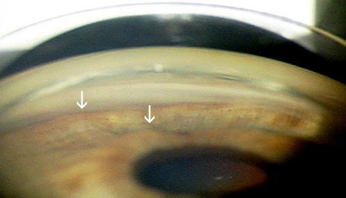
- A characteristic “double hump” sign can sometimes be seen on gonioscopy or ultrasound biomicroscopy, reflecting the iris draping over the anteriorly positioned ciliary processes
- Dark-room provocative testing or pharmacologic pupillary dilation can sometimes reproduce angle narrowing or pressure elevation despite the patent iridotomy, supporting the diagnosis
Diagnostic Imaging
Ultrasound biomicroscopy is the key imaging tool for confirming plateau iris configuration, directly visualizing the anteriorly positioned ciliary processes and their relationship to the peripheral iris and angle, findings not visible on standard anterior segment optical coherence tomography or slit-lamp examination alone given the need to image posterior to the iris.
Management
Confirming Iridotomy Patency First
Before attributing continued angle closure risk to plateau iris syndrome, confirming that the peripheral iridotomy itself is genuinely patent and adequately sized is an essential first step, since an inadequate iridotomy can produce a similar clinical picture of persistent risk despite an apparently completed laser procedure.
Laser Peripheral Iridoplasty
Argon laser peripheral iridoplasty, applying laser burns to the peripheral iris to contract the tissue and mechanically pull it away from the angle, directly addresses the angle-crowding mechanism specific to plateau iris and is a mainstay of treatment once the diagnosis is confirmed.
Medical Therapy
Topical miotics, such as pilocarpine, can help by pulling the iris away from the angle mechanically, though long-term use carries its own side effect considerations, and this approach is sometimes used as an adjunct or interim measure.
Lens Extraction
In some patients, particularly those with a coexisting cataract or with disease refractory to iridoplasty, lens extraction can help by creating additional anterior chamber space, indirectly reducing angle crowding, following similar reasoning to its use in other forms of angle closure.
Ongoing Monitoring
Because plateau iris syndrome reflects a persistent anatomic predisposition rather than a one-time, fully correctable problem, ongoing gonioscopic monitoring is appropriate even after initial successful treatment, since the underlying anteriorly positioned ciliary body configuration does not change and continued vigilance for recurrent angle narrowing is part of long-term care.
Prognosis
With correct recognition, distinguishing this condition from simple, iridotomy-responsive pupillary block, and with appropriate iridoplasty or other targeted treatment, most patients achieve good long-term angle stability and pressure control.
Missing the diagnosis, and assuming a patent iridotomy has fully addressed an eye’s angle closure risk when plateau iris configuration is actually the dominant mechanism, risks recurrent angle closure and optic nerve damage, which is why persistent risk after iridotomy should always prompt consideration of this specific, mechanistically distinct diagnosis.


Document what you see
Two smartphone imaging tools built for everyday clinic use — one for the slit lamp, one for the fundus.
From Choroida — the team behind this siteReferences
- Ritch R, Lowe RF. Angle-closure glaucoma: therapeutic overview. In: Ritch R, Shields MB, Krupin T, eds. The Glaucomas. 2nd ed. St. Louis: Mosby; 1996.
- Pavlin CJ, Ritch R, Foster FS. Ultrasound biomicroscopy in plateau iris syndrome. Am J Ophthalmol. 1992;113:390-395.
- Ritch R, Tham CC, Lam DS. Long-term success of argon laser peripheral iridoplasty in the management of plateau iris syndrome. Ophthalmology. 2004;111:104-108.
- Ritch R. Plateau iris is caused by abnormally positioned ciliary processes. J Glaucoma. 1992;1:23-26.
Test yourself
A few questions straight from this article.
-
In plateau iris, which structure crowds the anterior chamber angle?
The ciliary body sits unusually far anteriorly and pushes the peripheral iris against the trabecular meshwork, a mechanism iridotomy cannot address. -
What distinguishes plateau iris syndrome from plateau iris configuration?
Configuration describes the anatomy alone, while the syndrome is reserved for eyes that develop appositional or synechial closure despite a patent, functioning iridotomy. -
What is the central anterior chamber depth typically like in plateau iris syndrome?
The central chamber is often of relatively normal depth while the peripheral angle stays shallow, an important clue separating this from other angle closure causes. -
Which gonioscopic sign reflects the iris draping over anteriorly positioned ciliary processes?
A characteristic double hump sign can be seen on gonioscopy or ultrasound biomicroscopy as the iris drapes over the anteriorly placed ciliary processes. -
Which imaging modality confirms plateau iris configuration?
Ultrasound biomicroscopy directly visualises the anteriorly positioned ciliary processes and their relationship to the peripheral iris and angle. -
Why is ultrasound biomicroscopy preferred over anterior segment OCT in plateau iris?
The diagnosis depends on seeing the ciliary processes behind the iris, which standard anterior segment OCT and slit-lamp examination cannot reach. -
What is the essential first step before attributing persistent angle closure to plateau iris syndrome?
An inadequate iridotomy produces a similar picture of persistent risk, so its patency and size must be verified before the diagnosis is accepted. -
How does argon laser peripheral iridoplasty treat plateau iris syndrome?
Laser burns to the peripheral iris contract the tissue and mechanically pull it away from the angle, addressing the crowding mechanism directly. -
Which topical drug class helps in plateau iris syndrome by pulling the iris away from the angle?
Miotics pull the iris mechanically away from the angle and are used as an adjunct or interim measure, though long-term use brings side effect considerations. -
Why is ongoing gonioscopic monitoring needed after successful treatment of plateau iris syndrome?
The condition reflects a persistent anatomic predisposition rather than a one-time correctable problem, so vigilance for recurrent angle narrowing continues.
