
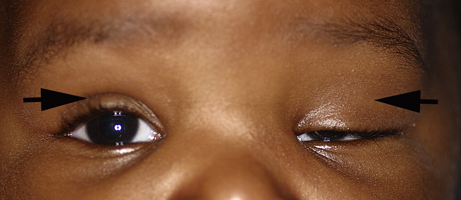
Pediatric blepharoptosis, or drooping of the upper eyelid in a child, is not simply a cosmetic finding, since in a developing visual system a drooping lid can directly cause amblyopia through two distinct mechanisms, either physically blocking the visual axis or inducing astigmatism from pressure on the developing cornea, making prompt assessment for amblyopia risk a central part of evaluating any child with ptosis.
Why Pediatric Ptosis Is Different From Adult Ptosis
In an adult, ptosis is primarily a functional and cosmetic concern, since the visual system is already mature and not at risk of amblyopia from the eyelid position itself.
In a child, particularly one within the sensitive period of visual development, the same drooping eyelid carries the additional risk of interfering with normal visual development, and the degree of this risk depends heavily on whether, and how much, the lid actually occludes the pupil and visual axis, and whether it is inducing significant astigmatism.
Mechanisms of Amblyopia in Ptosis
- Occlusion amblyopia: when the drooping lid margin covers a significant portion of the pupil, particularly the central visual axis, it functions similarly to patching, depriving that eye of adequate visual input during a critical developmental window
- Anisometropic or astigmatic amblyopia: even when the lid does not fully occlude the pupil, a ptotic eyelid can rest on and mechanically distort the underlying cornea, inducing astigmatism, which, if asymmetric between the two eyes, can itself cause amblyopia through anisometropia rather than direct visual axis blockage
All-fit Slit-Lamp Adapter
Record and share exactly what you see at the slit lamp. One adapter fits any slit lamp or surgical microscope — and any smartphone.
From Choroida — the team behind this siteCauses of Pediatric Ptosis
- Congenital myogenic ptosis, from a dystrophic, poorly developed levator muscle, the most common cause of pediatric ptosis
- Neurogenic causes, including congenital third nerve palsy and Horner syndrome
- Marcus Gunn jaw-winking ptosis, an unusual synkinetic pattern in which the eyelid elevates with jaw movement, from aberrant innervation between the trigeminal and levator muscles
- Mechanical ptosis, from an eyelid mass, such as a large chalazion or capillary hemangioma, weighing down the lid
- Blepharophimosis syndrome, a syndromic form of ptosis associated with other eyelid and periocular abnormalities
Assessment
- Measurement of the margin-reflex distance, quantifying the degree of ptosis, and assessment of how much of the pupil is covered in primary gaze
- Levator function testing, assessing how well the eyelid itself elevates, which is important both for understanding the underlying cause and for surgical planning
- Assessment of any compensatory chin-up head posture, which a child often adopts to keep the visual axis clear beneath a ptotic lid, and which itself is a sign that the ptosis is visually significant
- Cycloplegic refraction, to detect ptosis-induced astigmatism and any resulting anisometropia
- Assessment for amblyopia itself, through age-appropriate visual acuity testing, and monitoring for the development of a fixation preference favoring the non-ptotic eye
Management
Addressing Amblyopia Risk
When ptosis is visually significant, either from direct occlusion or induced astigmatism with anisometropia, standard amblyopia treatment, including correction of any refractive error and patching or other treatment of the amblyopia itself, proceeds alongside management of the ptosis, since delaying amblyopia treatment until after ptosis surgery can waste critical developmental time (see amblyopia).
Timing of Ptosis Surgery
- Severe, visually significant ptosis, particularly when it substantially occludes the visual axis, may need earlier surgical correction to remove the occlusive or astigmatism-inducing effect of the lid, even in infancy in select severe cases
- Milder ptosis, without significant amblyopia risk, is often observed and treated electively at an age when eyelid anatomy has matured further and surgical outcomes and cooperation are more favorable, often in the preschool years
- The decision balances the risk of amblyopia against the benefits of operating at an older age when levator function assessment and surgical planning are more reliable
Surgical Approach
Surgical technique depends on the underlying cause and measured levator function: eyes with good levator function are often managed with a levator resection or advancement, while eyes with poor levator function, as in severe congenital ptosis, more often require a frontalis sling procedure, using the frontalis muscle to elevate the lid via a sling material.
Long-Term Follow-Up
Children treated for ptosis need ongoing follow-up through the period of visual development, both to monitor for amblyopia recurrence or undertreatment and to assess for any recurrence of ptosis or need for surgical adjustment as the child grows.
Prognosis
With timely recognition of amblyopia risk and appropriate, coordinated management of both the ptosis and any resulting amblyopia, most children achieve good visual outcomes.
The greatest risk lies in ptosis that is not recognized as visually significant, allowing amblyopia to develop and progress unaddressed during the sensitive period of visual development.


Document what you see
Two smartphone imaging tools built for everyday clinic use — one for the slit lamp, one for the fundus.
From Choroida — the team behind this siteReferences
- Griepentrog GJ, Diehl NN, Mohney BG. Amblyopia in childhood eyelid ptosis. Am J Ophthalmol. 2013;155:1125-1128.
- Berry-Brincat A, Willshaw H. Paediatric blepharoptosis: a 10-year review. Eye (Lond). 2009;23:1554-1559.
- Anderson RL, Baumgartner SA. Amblyopia in ptosis. Arch Ophthalmol. 1980;98:1068-1069.
- Bagheri A, Borhani M, Salehirad S, Roodneshin F, Yazdani S. Amblyopia and its risk factors in patients with congenital ptosis. J Ophthalmic Vis Res. 2014;9:427-431.
Test yourself
A few questions straight from this article.
-
Why does blepharoptosis in a child carry risk beyond appearance?
In a developing visual system a drooping lid can directly cause amblyopia, so every child with ptosis needs prompt assessment of amblyopia risk. -
Pediatric ptosis can cause amblyopia by occlusion and through which second mechanism?
A ptotic lid resting on the cornea can distort it and induce astigmatism, which if asymmetric between eyes causes amblyopia through anisometropia. -
What is the most common cause of pediatric ptosis?
Congenital myogenic ptosis, caused by a dystrophic, poorly developed levator muscle, is the commonest cause in children. -
In Marcus Gunn jaw-winking ptosis, the eyelid elevates in response to which movement?
This synkinetic pattern arises from aberrant innervation, so the ptotic eyelid elevates when the jaw moves. -
Which measurement quantifies the degree of ptosis in a child?
Margin-reflex distance quantifies the ptosis, and it is assessed alongside how much of the pupil the lid covers in primary gaze. -
Which compensatory finding signals that a child's ptosis is visually significant?
Children adopt a chin-up posture to clear the visual axis beneath the drooping lid, and that posture is itself a sign the ptosis matters visually. -
Why is cycloplegic refraction performed in a child with blepharoptosis?
Cycloplegic refraction uncovers astigmatism induced by the lid and any resulting anisometropia, a route to amblyopia independent of axis occlusion. -
How is amblyopia treatment sequenced against ptosis surgery in a child with visually significant ptosis?
Refractive correction and patching proceed in parallel with ptosis management, because waiting until after lid surgery wastes critical developmental time. -
Which procedure is used for pediatric ptosis with poor levator function?
Poor levator function, as in severe congenital ptosis, usually calls for a frontalis sling, which lifts the lid using the frontalis muscle through sling material. -
Where does the greatest risk lie in pediatric blepharoptosis?
Most children do well with timely coordinated care, so the main danger is unrecognised visually significant ptosis letting amblyopia progress unaddressed.
