
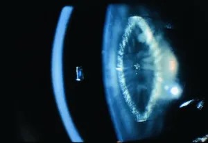
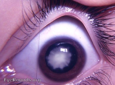
Zonular cataract, also called lamellar cataract, is the most common form of congenital and infantile cataract, characterized by opacification confined to a specific shell-like layer of the lens — usually surrounding a clear central nucleus and clear outer cortex — rather than diffusely affecting the entire lens.
The name comes from the appearance of the opacity as a discrete “zone” or lamella within the otherwise transparent lens, best visualized on retroillumination as a disc-shaped opacity with a clear center and clear periphery.
Pathogenesis and Associations
The opacity reflects a disturbance in lens fiber formation during a specific window of embryonic or early postnatal development, which is why it characteristically spares the lens fibers laid down before and after that window, producing the layered appearance.
It can be inherited (often autosomal dominant, sometimes with other family members affected in a similar pattern) or can arise sporadically.
It has been associated with a range of metabolic and systemic conditions, though many cases occur in otherwise healthy children with no identifiable cause.
Because the timing of the disturbance determines which specific lens layer is affected, the location and thickness of the opacity can, in a general sense, give a rough indication of when during development the causative insult or genetic influence occurred, even though this rarely changes management in an individual case.
Associated Conditions
- Hypocalcemia, particularly from hypoparathyroidism, one of the more classically taught metabolic associations
- Galactosemia and other inherited metabolic disorders affecting lens metabolism
- Maternal rubella infection during pregnancy, historically a significant cause before widespread vaccination
- Prematurity, independent of any specific identifiable metabolic cause
- Autosomal dominant inheritance as an isolated, non-syndromic familial trait
Because a meaningful proportion of cases are associated with a treatable systemic condition — hypocalcemia being the most immediately actionable — basic metabolic screening is a reasonable part of the initial workup in a newly identified zonular cataract, when bilateral or when other suggestive clinical features are present.
All-fit Slit-Lamp Adapter
Record and share exactly what you see at the slit lamp. One adapter fits any slit lamp or surgical microscope — and any smartphone.
From Choroida — the team behind this siteClinical Presentation
Visual impact varies considerably with the density and extent of the opacity: a thin, sparse lamellar opacity may cause minimal visual disturbance, while a dense, extensive one can significantly impair vision and carries a real risk of amblyopia if not identified and treated during the critical period of visual development in infancy and early childhood.
Riders — spoke-like radial opacities extending from the lamellar zone toward the lens equator — are sometimes seen as an additional feature and do not change the fundamental management approach.
Because a young infant cannot report symptoms directly, subtle behavioral clues — poor fixation and following, nystagmus, or a family’s report that the child does not seem to track faces as expected — are often what prompts the exam that first identifies the cataract, making a careful red reflex screen a standard part of every newborn and infant well-child visit.
Exam Findings
- A disc-shaped opacity in the deep cortex or perinuclear zone, sparing the central embryonic nucleus and the peripheral cortex
- Best appreciated on retroillumination, where it appears as a distinct ring or disc against the red reflex
- Often bilateral, though can be asymmetric in density
- Riders (radial spokes) in some cases
Differential Diagnosis
- Nuclear cataract (congenital form) — central opacity involving the embryonic and fetal nucleus, without the layered, disc-like pattern
- Anterior or posterior polar cataract — small, sharply localized to the anterior or posterior capsule rather than a deep, shell-like zone
- Total (complete) congenital cataract — diffuse opacification of the entire lens, generally denser and more visually significant from birth
- Sutural cataract — opacity confined to the Y-sutures of the lens, a distinct and generally more benign pattern
Evaluation
Beyond the ocular exam itself, a basic pediatric workup is reasonable for a newly diagnosed zonular cataract, especially bilateral cases: serum calcium and phosphate to screen for hypoparathyroidism, urine reducing substances or specific testing for galactosemia in the appropriate clinical context, and a review of maternal pregnancy history for rubella exposure or infection.
Family members should be examined when an inherited pattern is suspected, because identifying an affected but asymptomatic parent or sibling supports the diagnosis and informs genetic counseling.
Because untreated hypocalcemia and galactosemia both carry significant systemic risks well beyond the lens itself — including seizures in the former and severe, potentially life-threatening metabolic decompensation in the latter — the metabolic component of this workup should not be deferred or treated as an afterthought to the ophthalmic evaluation.
Management
Small, sparse, visually insignificant zonular cataracts can be monitored without intervention, with close attention to normal visual development and screening for any signs of amblyopia as the child grows.
Dense or extensive lesions causing significant visual impairment require surgical removal, ideally within the critical window for visual development, followed by appropriate optical correction (contact lens or, in older children, intraocular lens implantation) and amblyopia therapy as needed, with the exact timing of surgery balanced against the general risks of intraocular surgery in a very young infant.
Correcting any identified underlying metabolic cause — calcium replacement for hypoparathyroidism, dietary galactose restriction for galactosemia — is essential both for the child’s general health and, in some cases, to prevent further lens changes or fellow-eye involvement, even though it does not reverse opacity that has already formed.


Document what you see
Two smartphone imaging tools built for everyday clinic use — one for the slit lamp, one for the fundus.
From Choroida — the team behind this siteReferences
- Wilson ME, Trivedi RH, Pandey SK, eds. Pediatric Cataract Surgery: Techniques, Complications, and Management.
- Sheeladevi S, Lawrenson JG, Fielder AR, Suttle CM. Global prevalence of childhood cataract: a systematic review. Eye.
- American Academy of Ophthalmology. Basic and Clinical Science Course, Section 6: Pediatric Ophthalmology and Strabismus.
- American Academy of Ophthalmology. Basic and Clinical Science Course, Section 11: Lens and Cataract.
