
Climatic droplet keratopathy (also called Labrador keratopathy or spheroidal degeneration) is a degenerative corneal condition caused by chronic environmental exposure — ultraviolet radiation, wind, and dust — rather than any intrinsic corneal disease process.
It is most prevalent in populations with high occupational or recreational sun exposure at high altitude or high latitude with strong UV reflection off snow, water, or sand, in outdoor workers such as fishermen, farmers, and herders in these environments.
Because the condition develops slowly over decades of cumulative exposure, it is predominantly a disease of older adults in these high-exposure populations, and its severity in a given community tends to track closely with how much time residents have historically spent outdoors without effective eye protection.
Pathogenesis
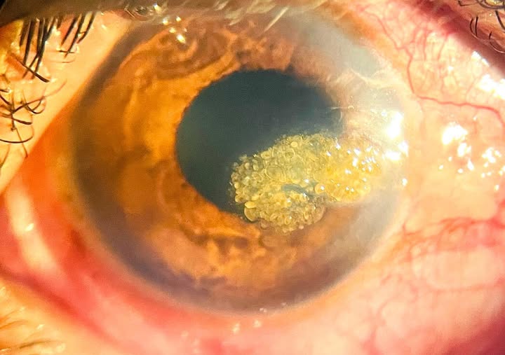
Chronic UV exposure damages corneal stromal proteins, which then aggregate and deposit as translucent, golden-brown spherules within the superficial to mid stroma, beginning in the interpalpebral zone — the horizontal band of cornea left exposed between the eyelids — where cumulative UV exposure is greatest.
Some evidence suggests the deposited material originates partly from degenerated, UV-damaged elastotic material similar to the changes seen in actinic skin damage, reflecting a shared underlying mechanism of chronic UV-induced tissue degeneration.
This shared mechanism with actinic skin changes is part of why the two so often coexist in the same patient, and why a weathered, sun-damaged periocular skin appearance in an older outdoor worker should prompt a careful slit-lamp look for early corneal spherules even before any visual complaint arises.
Clinical Presentation
Early disease is often asymptomatic or produces only mild irritation, and patients frequently have no complaints at all until the deposits become dense enough to affect vision, which usually takes many years of cumulative exposure to develop.
As the condition progresses, patients may notice glare, reduced vision, and a visible yellowish discoloration of the cornea that can become cosmetically apparent to others as well.
Glare and difficulty with night driving are often the earliest functional complaints reported, even before visual acuity on a standard chart has dropped meaningfully, because the scattered light from superficial deposits degrades contrast sensitivity and quality of vision before it reduces best-corrected acuity in a way easily captured by routine testing.
Because the disease affects an interpalpebral, outward-facing surface, many patients and even their families notice the cosmetic yellowing well before the patient reports any visual complaint at all, and this cosmetic concern — rather than any functional symptom — is what prompts a meaningful proportion of visits, particularly in patients whose occupation keeps them in regular contact with others who might comment on the appearance of their eyes.
All-fit Slit-Lamp Adapter
Record and share exactly what you see at the slit lamp. One adapter fits any slit lamp or surgical microscope — and any smartphone.
From Choroida — the team behind this siteExam Findings
- Golden-brown, translucent, droplet-like spherules in the superficial to mid corneal stroma
- Distribution concentrated in the interpalpebral zone, sparing the cornea protected by the eyelids
- Bilateral, though often asymmetric depending on differential sun exposure between the two eyes
- Advanced cases can show confluent, elevated nodular deposits significantly affecting the visual axis
- Often accompanied by other signs of chronic actinic exposure — pterygium, pinguecula, and actinic skin changes on the surrounding periocular skin
Staging
Climatic droplet keratopathy is generally staged from mild (fine, scattered peripheral deposits without visual impact) through moderate (denser, more central deposits with some visual symptoms) to severe (confluent, elevated deposits significantly obscuring vision).
Staging helps guide the threshold for intervention, because mild and even moderate disease is often managed conservatively while severe, vision-limiting disease may warrant surgical treatment.
Serial photography at each visit is a useful, low-cost way to document the stage and track any change over time, since the slow rate of progression means meaningful change is often only appreciable when comparing images taken years apart rather than at consecutive annual visits.
Differential Diagnosis
- Pinguecula and pterygium — related to the same UV exposure but represent conjunctival, not stromal corneal, degeneration
- Band keratopathy — calcium deposition, typically starts nasally and temporally rather than centrally, different composition and appearance
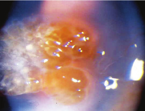
- Salzmann’s nodular degeneration — bluish-white, elevated nodules, often following chronic ocular surface inflammation rather than UV exposure specifically
- Lattice or granular corneal dystrophy — inherited, bilateral, without the characteristic environmental exposure history or interpalpebral distribution
The occupational and geographic exposure history, together with the classic interpalpebral distribution and coexisting pterygium/pinguecula, usually makes the diagnosis straightforward without needing further testing.
Management
Prevention through UV-blocking sunglasses and broad-brimmed hats is the most effective intervention and should be emphasized early in populations at occupational or environmental risk, because established deposits do not regress and the goal shifts to preventing further accumulation.
Mild disease requires no treatment beyond ongoing UV protection and monitoring.
For moderate to severe disease affecting vision, superficial keratectomy (manual or with phototherapeutic keratectomy using an excimer laser) can remove the more superficial deposits and improve vision, though deeper deposits may not be fully accessible to superficial techniques.
Lamellar or, in advanced cases, penetrating keratoplasty is reserved for severe, vision-limiting disease with deep stromal involvement not amenable to superficial removal.
Regardless of the treatment chosen, ongoing UV protection remains essential afterward, since the same environmental exposure that produced the original deposits can drive recurrence within a surgically treated or transplanted cornea if sun protection habits are not maintained going forward, making patient education about this point just as important as the procedure itself.


Document what you see
Two smartphone imaging tools built for everyday clinic use — one for the slit lamp, one for the fundus.
From Choroida — the team behind this siteReferences
- Norn MS. Spheroid degeneration of the cornea and conjunctiva (Labrador keratopathy). Acta Ophthalmologica.
- Gray RH, Johnson GJ, Freedman A. Climatic droplet keratopathy. Survey of Ophthalmology.
- Freedman A, Gray RH. Climatic droplet keratopathy in an Israeli Bedouin population. British Journal of Ophthalmology.
- American Academy of Ophthalmology. Basic and Clinical Science Course, Section 8: External Disease and Cornea.
