
Bull’s-Eye Maculopathy is a description, not a diagnosis. The concentric ring of parafoveal atrophy around a spared fovea is instantly recognisable, and the reflex is to blame hydroxychloroquine.

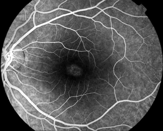
But a target-shaped macula has many causes, and several are inherited diseases that no drug review will explain.
For clinicians, recognizing that Bull’s-Eye Maculopathy is a pattern with a broad differential matters because the work-up and the counselling differ completely between a drug toxicity and a cone dystrophy.
The task is not to name the ring, but to find out what made it.
What Is Bull’s-Eye Maculopathy?
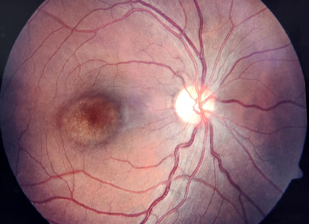
Bull’s-Eye Maculopathy is a ring of parafoveal retinal pigment epithelial depigmentation and atrophy that surrounds a relatively spared central fovea, giving a target appearance.
The key points are:
- It is a morphological sign, not a single disease
- The fovea is characteristically spared, at least early
- It reflects loss of the parafoveal photoreceptors and RPE
- Its causes span toxic, hereditary, and degenerative disease
Seeing the ring should trigger a differential, not a diagnosis.
Causes and Differential Diagnosis
The differential is the heart of the topic.
- Hydroxychloroquine and chloroquine toxicity — the classic drug cause, dose- and duration-related
- Cone and cone-rod dystrophy — photophobia, poor colour vision, abnormal cone responses on ERG
- Stargardt disease — flecks and a dark choroid on angiography
- Age-related macular degeneration and central areolar choroidal dystrophy
- Benign concentric annular macular dystrophy
- Systemic and neurodegenerative disease — Bardet–Biedl syndrome and Batten disease
- Other drugs — clofazimine and, rarely, others
Age, drug history, symptoms, and family history are what separate these causes long before any test is ordered.
Why the Hydroxychloroquine Reflex Misleads
Attributing every bull’s-eye to hydroxychloroquine causes errors in both directions.
- A young patient with photophobia and poor colour vision is far more likely to have a cone dystrophy than drug toxicity
- Labelling an inherited dystrophy as “drug toxicity” delays genetic diagnosis and counselling
- Conversely, dismissing true early hydroxychloroquine damage misses a chance to stop the drug
The drug history must fit the dose, duration, and imaging before hydroxychloroquine is accepted as the cause.
Diagnostic Evaluation
Multimodal imaging and electrophysiology sort the causes out.
Imaging
- Fundus autofluorescence shows a parafoveal ring of altered autofluorescence
- OCT demonstrates parafoveal loss of the ellipsoid zone and outer retina, with foveal sparing
Functional and Genetic Testing
- Electroretinography distinguishes a generalised cone or cone-rod dystrophy from a localised toxicity
- Genetic testing confirms an inherited dystrophy and guides counselling
- A careful drug and dosing history remains essential throughout
Imaging shows the ring; electrophysiology and genetics reveal why it is there.
Management
Management depends entirely on the underlying cause.
- Drug toxicity — stop or review the offending agent in discussion with the prescriber
- Inherited dystrophies — genetic counselling, low-vision support, and monitoring
- Age-related and degenerative causes — managed on their own pathways
- All patients benefit from documentation and structured follow-up
The correct treatment starts with the correct cause, which is why the differential comes first.
Prognosis
Outcome tracks the underlying disease.
- Early drug toxicity may stabilise once the drug is stopped, though damage can progress
- Inherited dystrophies are typically progressive
- Foveal involvement, whatever the cause, marks a worse visual prognosis
In practice, the value of the bull’s-eye sign is that it forces a search for a specific, sometimes treatable, cause.
Would you have interest in taking retinal images with your smartphone?
Fundus photography lets you document and monitor a bull’s-eye macular lesion over time and share the images with colleagues and patients.
RETINAL IMAGING BY YOUR SMARTPHONE
References
- Marmor MF, Kellner U, Lai TYY, Melles RB, Mieler WF. “Recommendations on Screening for Chloroquine and Hydroxychloroquine Retinopathy (2016 Revision).” Ophthalmology. 2016;123(6):1386–1394.
- Kurz-Levin MM, Halfyard AS, Bunce C, et al. “Clinical Variations in Assessment of Bull’s-Eye Maculopathy.” Archives of Ophthalmology. 2002;120(5):567–575.
- Michaelides M, Hardcastle AJ, Hunt DM, Moore AT. “Progressive Cone and Cone-Rod Dystrophies: Phenotypes and Underlying Molecular Genetic Basis.” Survey of Ophthalmology. 2006;51(3):232–258.
- American Academy of Ophthalmology. “Bull’s Eye Maculopathy.” EyeWiki.

