
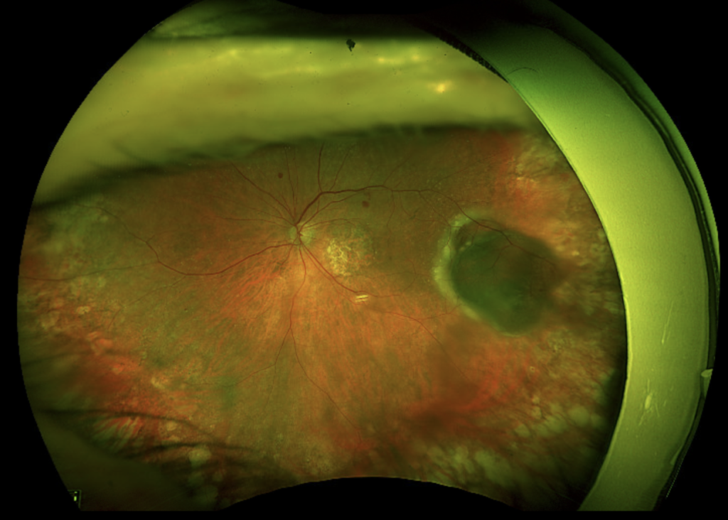
Peripheral Exudative Hemorrhagic Chorioretinopathy is the reason some healthy eyes are nearly removed for a cancer that was never there.
It produces a dark, elevated mass in the far periphery of the fundus, packed with blood and lipid, that looks unnervingly like a choroidal melanoma.
Yet it is a degenerative, self-limiting condition of the elderly retina, not a tumour.
For clinicians, recognizing Peripheral Exudative Hemorrhagic Chorioretinopathy (PEHCR) matters because the alternative—an unnecessary enucleation—is catastrophic and avoidable.
Any peripheral haemorrhagic lesion in an older patient deserves the question: is this a melanoma, or is it PEHCR?
What Is Peripheral Exudative Hemorrhagic Chorioretinopathy?
PEHCR is a degenerative disorder of the peripheral retina and choroid characterized by sub-retinal and sub-RPE haemorrhage and exudation.
Its defining features are:
- An elevated, often dark peripheral lesion, usually temporal
- Sub-retinal or sub-RPE haemorrhage with lipid exudation
- A strong association with age-related macular degeneration
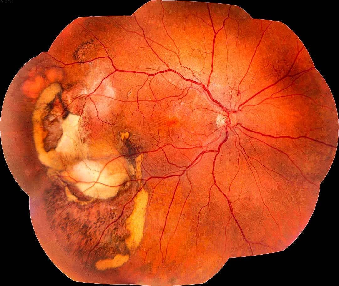
- A tendency to resolve, leaving pigmentary and fibrotic scarring
It is best understood as a peripheral counterpart of neovascular AMD, driven by abnormal choroidal vessels far from the macula.
Epidemiology
The condition is a disease of the older eye.
- Typically affects patients in their seventies and eighties
- More common in women
- Bilateral in roughly a third of cases
- Frequently associated with macular AMD in the same or fellow eye
Because the lesions sit in the far periphery, many are found incidentally on routine or widefield examination.
Fundus Explorer Pro
Photograph the retinal findings described here with the phone already in your pocket — 22 D optics and built-in illumination in one handheld unit.
From Choroida — the team behind this sitePathophysiology
The mechanism parallels that of neovascular macular disease.
- Abnormal peripheral choroidal neovascular or polypoidal vessels develop, often on a background of RPE and Bruch membrane degeneration
- These fragile vessels leak and bleed beneath the RPE and retina
- Blood and lipid accumulate to form an elevated, tumour-like lesion
- Over time the haemorrhage organizes into fibrosis and pigment
It is this combination of elevation, dark blood, and lipid that so convincingly imitates a pigmented tumour.
Clinical Presentation
Symptoms
Most patients have few or no symptoms.
- Often completely asymptomatic and found incidentally
- Painless visual loss if haemorrhage or exudate reaches the macula
- Floaters or sudden loss if the lesion bleeds into the vitreous
Examination Findings
The appearance is dramatic but characteristic.
- An elevated peripheral lesion, usually temporal, with dark sub-retinal blood
- Lipid exudation and sub-retinal fluid around the lesion
- Sub-RPE haemorrhage that may appear almost black
- Associated drusen and RPE changes at the macula
- Occasionally vitreous haemorrhage obscuring the view
A peripheral haemorrhagic lesion with lipid, in an elderly eye that also shows macular AMD, is far more likely to be PEHCR than melanoma.
Why PEHCR Is So Often Mistaken for Melanoma
The confusion is understandable and follows a familiar pattern.
- The lesion is elevated, dark, and dome-shaped, exactly like a pigmented tumour
- It sits in the periphery, where it is hard to examine and image
- Sub-RPE blood can look solid and mass-like on cursory examination
- Fear of missing a melanoma pushes clinicians toward aggressive assumptions
In a large series, a substantial number of eyes referred as suspected choroidal melanoma were in fact PEHCR—underlining how often the two are confused.
Diagnostic Evaluation
Imaging is what separates a benign haemorrhage from a true tumour.
Ultrasonography
- B-scan typically shows haemorrhage and a dome of sub-RPE blood rather than a solid, acoustically hollow mass
- Melanoma classically shows low internal reflectivity and choroidal excavation, which PEHCR lacks
Angiography and OCT
- Fluorescein and indocyanine green angiography may reveal peripheral polypoidal or neovascular lesions
- Blocked fluorescence from blood, rather than a vascularized tumour, supports PEHCR
- OCT of the lesion, where reachable, shows haemorrhagic RPE detachment
The combination of a haemorrhagic, non-solid lesion on ultrasound and bilateral peripheral disease strongly favours PEHCR.
Differential Diagnosis
Several elevated peripheral lesions enter the differential:
- Choroidal melanoma — the critical distinction, favouring a solid, acoustically hollow mass
- Choroidal melanocytoma and other pigmented lesions
- Choroidal metastasis and choroidal haemangioma
- Retinal vasoproliferative tumour
- Peripheral neovascular AMD and Coats disease
Bilaterality, coexisting macular AMD, and haemorrhage without a solid mass are the features that point away from melanoma.
Management
Most lesions need confidence rather than intervention.
- Observation is appropriate for asymptomatic peripheral lesions that are not threatening the macula
- Intravitreal anti-VEGF is used when there is macular exudation or fluid
- Laser photocoagulation or cryotherapy can treat active peripheral leakage
- Vitrectomy is reserved for non-clearing vitreous haemorrhage
- The essential step is to confirm the diagnosis and avoid enucleation
Once melanoma has been excluded, the natural tendency of PEHCR to resolve makes conservative management the rule.
Prognosis
The outlook is generally favourable.
- Peripheral lesions often resolve with pigmentary and fibrotic scarring
- Central vision is preserved unless the macula is involved
- The main threat is diagnostic—an eye lost to a mistaken cancer diagnosis
In practice, the whole value of knowing PEHCR is protecting a comfortable, seeing eye from unnecessary removal.


Document what you see
Two smartphone imaging tools built for everyday clinic use — one for the slit lamp, one for the fundus.
From Choroida — the team behind this siteReferences
- Shields CL, Salazar PF, Mashayekhi A, Shields JA. “Peripheral Exudative Hemorrhagic Chorioretinopathy Simulating Choroidal Melanoma in 173 Cases.” Ophthalmology. 2009;116(3):529–535.
- Annesley WH Jr. “Peripheral Exudative Hemorrhagic Chorioretinopathy.” Transactions of the American Ophthalmological Society. 1980;78:321–364.
- Mantel I, Uffer S, Zografos L. “Peripheral Exudative Hemorrhagic Chorioretinopathy: A Clinical, Angiographic, and Histologic Study.” American Journal of Ophthalmology. 2009;148(6):932–938.
- Reese AB, Jones IS. “Hematomas Under the Retinal Pigment Epithelium.” Transactions of the American Ophthalmological Society. 1962;60:43–79.
- American Academy of Ophthalmology. “Peripheral Exudative Hemorrhagic Chorioretinopathy.” EyeWiki.
Test yourself
A few questions straight from this article.
-
Peripheral exudative hemorrhagic chorioretinopathy (PEHCR) is best defined as which kind of disorder?
PEHCR is a degenerative disorder of the peripheral retina and choroid marked by sub-retinal and sub-RPE haemorrhage with lipid exudation, not a tumour. -
Which macular disease is strongly associated with peripheral exudative hemorrhagic chorioretinopathy?
PEHCR behaves as a peripheral counterpart of neovascular AMD and is frequently accompanied by macular AMD in the same or the fellow eye. -
Which patient group is typically affected by peripheral exudative hemorrhagic chorioretinopathy?
PEHCR is a disease of the older eye, usually presenting in the seventies and eighties and occurring more commonly in women. -
In roughly what proportion of patients with PEHCR is the disease bilateral?
PEHCR is bilateral in roughly a third of cases, and bilaterality is one of the features that argues against choroidal melanoma. -
What is the underlying mechanism of the lesion in peripheral exudative hemorrhagic chorioretinopathy?
Fragile peripheral choroidal neovascular or polypoidal vessels arise on degenerate RPE and Bruch membrane, then bleed and leak beneath the RPE and retina. -
Where in the fundus does the lesion of PEHCR most characteristically sit?
The classic PEHCR lesion is an elevated, dark, usually temporal peripheral mass of sub-retinal and sub-RPE blood with surrounding lipid. -
Which B-scan ultrasound picture supports PEHCR rather than choroidal melanoma?
PEHCR shows haemorrhage and a dome of sub-RPE blood, while melanoma classically shows low internal reflectivity and choroidal excavation that PEHCR lacks. -
How should an asymptomatic peripheral PEHCR lesion that spares the macula be managed?
Asymptomatic peripheral lesions that do not threaten the macula are observed; PEHCR tends to resolve, and the essential step is confirming the diagnosis to avoid enucleation. -
Which treatment is used in PEHCR when there is macular exudation or subretinal fluid?
Intravitreal anti-VEGF is used when exudation or fluid reaches the macula, while laser or cryotherapy targets active peripheral leakage. -
What is the principal threat to the eye in peripheral exudative hemorrhagic chorioretinopathy?
PEHCR usually resolves with pigmentary and fibrotic scarring and spares central vision; the real danger is diagnostic, losing a comfortable seeing eye to a cancer that was never there.

Writes and reviews the clinical articles on Educate. Every post is checked against peer-reviewed sources before it goes live.


