
Introduction
Despite significant advances in antimicrobial therapy and diagnostic tools, microbial keratitis remains a major cause of visual morbidity worldwide.

While many cases respond well to standard treatment protocols, a subset of patients demonstrates poor or delayed response, often leading to corneal scarring, perforation, or even loss of the eye.
Understanding why some keratitis cases fail treatment is critical—not only for improving outcomes but also for refining clinical judgment in challenging scenarios.
This article explores the often-overlooked pitfalls that contribute to treatment failure in keratitis, with a focus on practical, real-world clinical insights.
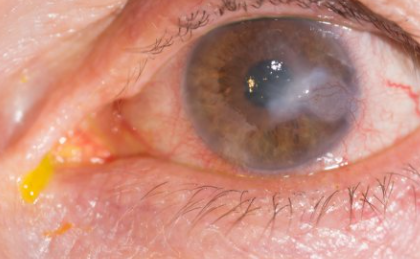
1. Misdiagnosis: The Most Common Trap
One of the leading causes of treatment failure is incorrect initial diagnosis. Not all corneal ulcers are bacterial, yet many are treated empirically as such.
Common Misdiagnoses:
- Fungal keratitis mistaken for bacterial keratitis
- Acanthamoeba keratitis misdiagnosed as herpetic keratitis
- Sterile inflammatory keratitis is treated as infectious
Why This Matters:
Each type of keratitis requires a completely different treatment approach. For example:
- Fungal keratitis requires antifungal agents, not antibiotics
- Acanthamoeba requires prolonged anti-protozoal therapy
- Herpetic keratitis worsens with steroids if epithelial disease is present
👉 Clinical Tip: If there is no improvement within 48–72 hours of appropriate therapy, reconsider the diagnosis immediately.
2. Delayed or Inadequate Microbiological Workup
Empirical treatment is common, but failure to perform corneal scraping and culture can lead to inappropriate therapy.
Pitfalls:
- Skipping microbiology in “small” ulcers
- Prior antibiotic use masks culture results
- Inadequate sample collection
Consequences:
- Missed atypical organisms
- Inability to tailor therapy
- Increased resistance risk
👉 Clinical Tip: Always consider microbiological investigation in:
- Central ulcers
- Large or deep infiltrates
- Non-responding cases
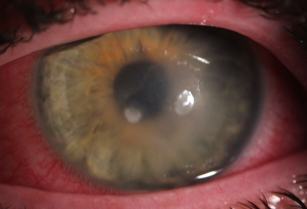
3. Drug Penetration and Pharmacokinetics
Even when the correct drug is chosen, poor corneal penetration may limit effectiveness.
Contributing Factors:
- Intact epithelium limits drug entry
- Deep stromal or endothelial infections
- Biofilm formation (especially in contact lens-related infections)
Example:
Topical antifungals like natamycin have limited stromal penetration, making deep fungal infections difficult to treat.
👉 Clinical Tip: Consider:
- Debridement to enhance penetration
- Switching to fortified or systemic therapy in deep infections
4. Patient Compliance Issues
Non-adherence to treatment regimens is an underestimated factor.
Challenges:
- Frequent dosing schedules (every 30–60 minutes)
- Long treatment duration
- Financial constraints
- Lack of patient education
Impact:
Even the most appropriate treatment will fail if not followed correctly.
👉 Clinical Tip:
Simplify regimens when possible and ensure patients understand:
- Severity of condition
- Importance of adherence
- Warning signs of deterioration
5. Steroid Misuse
Topical steroids can be a double-edged sword.
Common Errors:
- Early use before infection control
- Use in undiagnosed keratitis
- Continued use despite worsening infection
Risks:
- Enhanced microbial replication
- Masking clinical signs
- Corneal thinning and perforation
👉 Clinical Tip:
Avoid steroids in active epithelial keratitis and use them cautiously only after the infection is under control.
6. Resistant and Atypical Organisms
Emerging antimicrobial resistance is a growing concern.
Examples:
- Methicillin-resistant Staphylococcus aureus (MRSA)
- Multidrug-resistant Pseudomonas
- Rare organisms like Nocardia or Mycobacteria
Clinical Clues:
- Poor response to broad-spectrum antibiotics
- Indolent or atypical clinical course
👉 Clinical Tip:
Reassess and escalate treatment early in non-responding cases. Consider referral if needed.
7. Underlying Ocular Surface Disease
Pre-existing conditions often interfere with healing.
Common Factors:
- Dry eye disease
- Neurotrophic keratopathy
- Exposure keratopathy
- Lid abnormalities
Impact:
- Delayed epithelial healing
- Increased susceptibility to infection
- Higher recurrence rates
👉 Clinical Tip:
Always treat the underlying surface disease, not just the infection.
8. Contact Lens-Related Factors
Contact lens wear remains a major risk factor.
Issues:
- Poor hygiene
- Extended wear
- Contaminated solutions
Special Concern:
- High risk of Acanthamoeba keratitis
- Biofilm-associated infections
👉 Clinical Tip:
Always ask detailed history about lens use—even if the patient doesn’t volunteer it.
9. Late Presentation
In many settings, patients present late after:
- Self-medication
- Traditional remedies
- Delayed referral
Consequences:
- Advanced stromal involvement
- Increased risk of perforation
- Limited treatment options
👉 Clinical Tip:
Early referral and aggressive management are key to preserving vision.
Conclusion
Failure of keratitis treatment is rarely due to a single factor. Instead, it is usually a combination of diagnostic errors, inadequate therapy, patient-related factors, and underlying ocular conditions.
Recognizing these hidden pitfalls can significantly improve clinical outcomes.
For ophthalmologists, the key lies in:
- Maintaining a high index of suspicion
- Reassessing non-responding cases early
- Adopting a multidisciplinary and individualized approach
Ultimately, timely intervention and critical thinking—not just protocols—are what save vision.
References
- Ting DSJ, et al. “Infectious keratitis: an update on epidemiology, causative microorganisms, risk factors, and antimicrobial resistance.” Eye (Lond). 2021.
- Austin A, et al. “Microbial keratitis: diagnosis and management.” Ophthalmology. 2017.
- Thomas PA, Kaliamurthy J. “Mycotic keratitis: epidemiology, diagnosis and management.” Clin Microbiol Infect. 2013.
- Stapleton F, et al. “The epidemiology of microbial keratitis with contact lens use.” Eye. 2012.
- Sharma N, et al. “Evaluation of corneal scraping smear examination in the diagnosis of microbial keratitis.” Br J Ophthalmol. 2010.
- Alfonso EC, et al. “Fungal keratitis.” Lancet. 2019.
- Dart JK, et al. “Acanthamoeba keratitis: diagnosis and treatment update.” Am J Ophthalmol. 2009.

RETINAL IMAGING BY YOUR SMARTPHONE