
Case Study
A 46-year-old woman presented for routine ophthalmic evaluation with mild complaints of intermittent blurred central vision in both eyes.
She reported no history of night blindness or significant visual loss. Her medical history was unremarkable. Best-corrected visual acuity was 20/25 in both eyes.
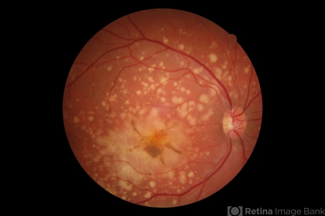
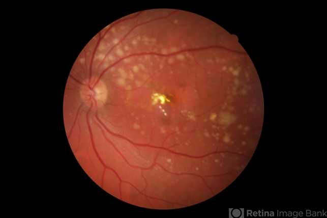
Anterior segment examination was normal. Fundus examination revealed bilateral, symmetric pigmentary changes in the macular region with a distinctive butterfly-shaped pattern radiating from the fovea.
The lesions consisted of irregular, yellow-brown pigment deposition at the level of the retinal pigment epithelium (RPE).
Optical coherence tomography (OCT) demonstrated subtle irregularities and hyperreflective deposits at the RPE level without significant disruption of the outer retinal layers.
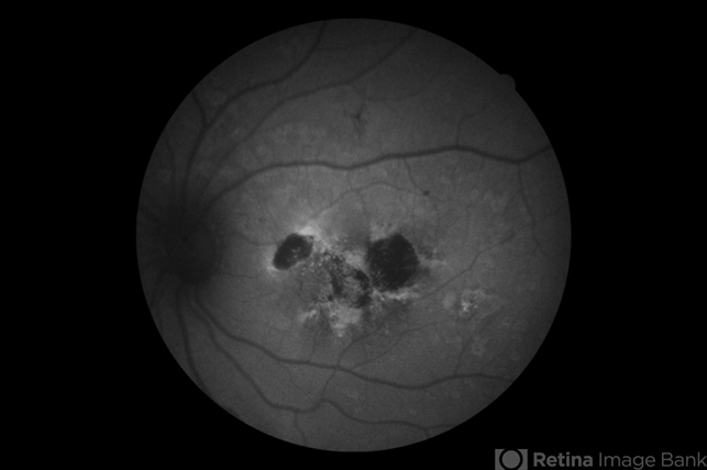
Fundus autofluorescence (FAF) showed a characteristic pattern of mixed hyperautofluorescent and hypoautofluorescent areas corresponding to the pigmentary changes.
Full-field electroretinography (ERG) was normal. Based on the clinical findings and characteristic macular appearance, a diagnosis of butterfly-shaped pattern dystrophy was established.
Disease Entity
Butterfly-shaped pattern dystrophy is a form of pattern dystrophy of the retinal pigment epithelium characterized by pigmentary deposits in the macula arranged in a butterfly-like configuration.
It belongs to a group of inherited macular disorders collectively referred to as pattern dystrophies.
Pattern dystrophies are typically associated with mutations in the peripherin/RDS (PRPH2) gene, which encodes a photoreceptor membrane glycoprotein essential for maintaining the structural integrity of photoreceptor outer segments.
Butterfly-shaped pattern dystrophy is characterized by the accumulation of lipofuscin-like material within the RPE, producing distinctive pigmentary patterns visible on fundus examination.
The disease usually manifests in mid-adulthood and generally follows a relatively benign course compared with other macular dystrophies.
Fundus Explorer Pro
Photograph the retinal findings described here with the phone already in your pocket — 22 D optics and built-in illumination in one handheld unit.
From Choroida — the team behind this sitePathophysiology
The pathogenesis of butterfly-shaped pattern dystrophy involves dysfunction of the retinal pigment epithelium and abnormal metabolism of photoreceptor outer segments.
Mutations in the PRPH2 gene disrupt the normal structure and function of photoreceptor outer segment discs.
This leads to impaired processing of photoreceptor debris by the RPE and subsequent accumulation of lipofuscin and other metabolic byproducts.
Lipofuscin accumulation within RPE cells alters their normal metabolic function and produces characteristic pigmentary changes visible clinically.
The distribution of these deposits in a butterfly-shaped pattern forms radiating patterns resembling butterfly wings centered on the macula.
Despite these structural abnormalities, photoreceptor function often remains relatively preserved for many years, which explains the mild symptoms and slow disease progression.
Over time, however, progressive RPE dysfunction may lead to complications such as geographic atrophy or choroidal neovascularization.
Epidemiology
Butterfly-shaped pattern dystrophy is a rare inherited retinal disease. It most commonly presents in individuals between the third and fifth decades of life.
The disorder typically follows an autosomal dominant inheritance pattern, although sporadic cases may occur.
Both sexes are affected equally, and there is no known racial predilection.
Because symptoms are often mild, the condition may remain undiagnosed until middle age when routine ophthalmic examinations reveal characteristic fundus changes.
Clinical Features
Many patients with butterfly-shaped pattern dystrophy are asymptomatic at early stages.
When symptoms occur, they may include:
-
Mild central visual blur
-
Metamorphopsia
-
Slight reduction in visual acuity
-
Difficulty reading in dim illumination
Visual acuity is often preserved in early stages, frequently remaining better than 20/40.
The hallmark clinical feature is a bilateral, symmetric pigmentary pattern in the macula resembling butterfly wings.
These pigment deposits appear as:
-
Yellow-brown or grayish lesions
-
Radiating from the foveal center
-
Located at the level of the RPE
The lesions may become more prominent over time.
Examination Findings
Anterior segment examination is usually normal.
Fundus examination reveals:
-
Characteristic butterfly-shaped pigment deposition
-
Macular involvement centered around the fovea
-
Symmetric bilateral lesions
-
Minimal surrounding retinal changes in early stages
Optical Coherence Tomography (OCT)
OCT findings may include:
-
Hyperreflective deposits at the level of the RPE
-
Mild irregularity of the RPE band
-
Preserved ellipsoid zone in early disease
Fundus Autofluorescence (FAF)
FAF typically demonstrates:
-
Areas of hyperautofluorescence corresponding to lipofuscin accumulation
-
Patchy hypoautofluorescence in regions of RPE degeneration
Fluorescein Angiography (FA)
FA may show:
-
Window defects due to RPE changes
-
Minimal leakage in most cases
Electrophysiologic Testing
Full-field ERG is usually normal. Multifocal ERG may show localized macular dysfunction in advanced stages.
Differential Diagnosis
Butterfly-shaped pattern dystrophy should be distinguished from other macular disorders with pigmentary changes.
Important differential diagnoses include:
-
Adult-onset foveomacular vitelliform dystrophy
-
Stargardt disease
-
Age-related macular degeneration (AMD)
-
Best vitelliform macular dystrophy
-
Other pattern dystrophies of the RPE
Stargardt disease typically presents earlier in life and is associated with widespread flecks and progressive visual loss.
Age-related macular degeneration generally occurs later in life and is associated with drusen and geographic atrophy.
Best disease shows characteristic vitelliform lesions and abnormal electro-oculography.
Careful clinical evaluation and imaging help distinguish these conditions.
Diagnosis
Diagnosis is based on characteristic clinical findings and supportive imaging studies.
Diagnostic features include:
-
Butterfly-shaped pigment pattern in the macula
-
Bilateral symmetric lesions
-
Onset in mid-adulthood
-
Normal or near-normal ERG
Genetic testing may confirm PRPH2 mutations, although it is not always required.
Multimodal imaging, including OCT and FAF, plays an important role in confirming the diagnosis and monitoring disease progression.
Management
There is currently no specific treatment for butterfly-shaped pattern dystrophy.
Management focuses on monitoring for potential complications, particularly:
-
Choroidal neovascularization
-
Progressive macular atrophy
Recommended management strategies include:
-
Regular ophthalmic examinations
-
Periodic OCT imaging
-
Patient education regarding symptoms of neovascular complications
If choroidal neovascularization develops, treatment with intravitreal anti–vascular endothelial growth factor (anti-VEGF) agents may be indicated.
Low-vision aids may be useful in advanced cases with significant central vision impairment.
Prognosis
The prognosis for butterfly-shaped pattern dystrophy is generally favorable.
Most patients maintain relatively good visual acuity for many years, and disease progression is usually slow.
However, some individuals may develop complications such as:
-
Choroidal neovascularization
-
Geographic atrophy of the RPE
-
Progressive central vision loss
Regular monitoring is therefore recommended.


Document what you see
Two smartphone imaging tools built for everyday clinic use — one for the slit lamp, one for the fundus.
From Choroida — the team behind this siteReferences
-
American Academy of Ophthalmology. Basic and Clinical Science Course (BCSC): Retina and Vitreous. San Francisco, CA: AAO; latest edition.
-
Yanoff M, Duker JS. Ophthalmology. 5th ed. Elsevier; 2019.
-
Traboulsi EI. Genetic Diseases of the Eye. 2nd ed. Oxford University Press, 2012.
-
Marmor MF, Byers B. Pattern dystrophy of the retinal pigment epithelium. Arch Ophthalmol. 1977;95(4):646–652.
-
Boon CJ, van Schooneveld MJ, den Hollander AI, et al. The spectrum of retinal dystrophies caused by mutations in the peripherin/RDS gene. Prog Retin Eye Res. 2008;27(2):213–235.
-
Querques G, Massamba N, Srour M, et al. Pattern dystrophies of the retinal pigment epithelium: a review. Surv Ophthalmol. 2011;56(1):1–27.
Test yourself
A few questions straight from this article.
-
Mutations in which gene are typically associated with butterfly-shaped pattern dystrophy?
Pattern dystrophies are typically caused by PRPH2 mutations, which disrupt the photoreceptor outer segment discs and impair RPE processing of photoreceptor debris. -
Accumulation of which material in the RPE produces the pigmentary changes of butterfly-shaped pattern dystrophy?
Impaired handling of photoreceptor debris leads to lipofuscin accumulation inside RPE cells, producing the clinically visible pigmentary pattern. -
In which decades of life does butterfly-shaped pattern dystrophy most commonly present?
The condition usually manifests in mid-adulthood, most often between the third and fifth decades, and mild symptoms mean it is often found incidentally. -
What is the expected full-field electroretinogram result in butterfly-shaped pattern dystrophy?
Full-field ERG is usually normal because generalized retinal function is preserved; multifocal ERG may show localized macular dysfunction in advanced disease. -
What is the hallmark fundus finding in butterfly-shaped pattern dystrophy?
The hallmark is bilateral, symmetric yellow-brown pigment at the RPE level radiating from the foveal centre in a butterfly-wing configuration. -
What does fundus autofluorescence typically show in butterfly-shaped pattern dystrophy?
FAF shows hyperautofluorescence where lipofuscin has accumulated and patchy hypoautofluorescence where the RPE has degenerated. -
Which finding points to Best disease rather than butterfly-shaped pattern dystrophy?
Best vitelliform macular dystrophy shows a characteristic vitelliform lesion together with abnormal electro-oculography, unlike pattern dystrophy. -
What visual acuity is typically preserved in early butterfly-shaped pattern dystrophy?
Photoreceptor function stays relatively preserved for years, so acuity in early disease is often better than 20/40 despite striking fundus changes. -
Which complication of butterfly-shaped pattern dystrophy is treated with intravitreal anti-VEGF therapy?
No specific treatment exists for the dystrophy, but if choroidal neovascularization develops, intravitreal anti-VEGF agents may be indicated. -
Which inheritance pattern does butterfly-shaped pattern dystrophy usually follow?
The disorder typically follows autosomal dominant inheritance, although sporadic cases occur; both sexes are affected equally.
