
Case Study
A 62-year-old man with a history of uncontrolled diabetes and chronic kidney disease presented with a sudden drop in vision and mild eye pain in his left eye for four days.

He also reported fever and lethargy for the past week. His best-corrected visual acuity was counting fingers at 1 meter.
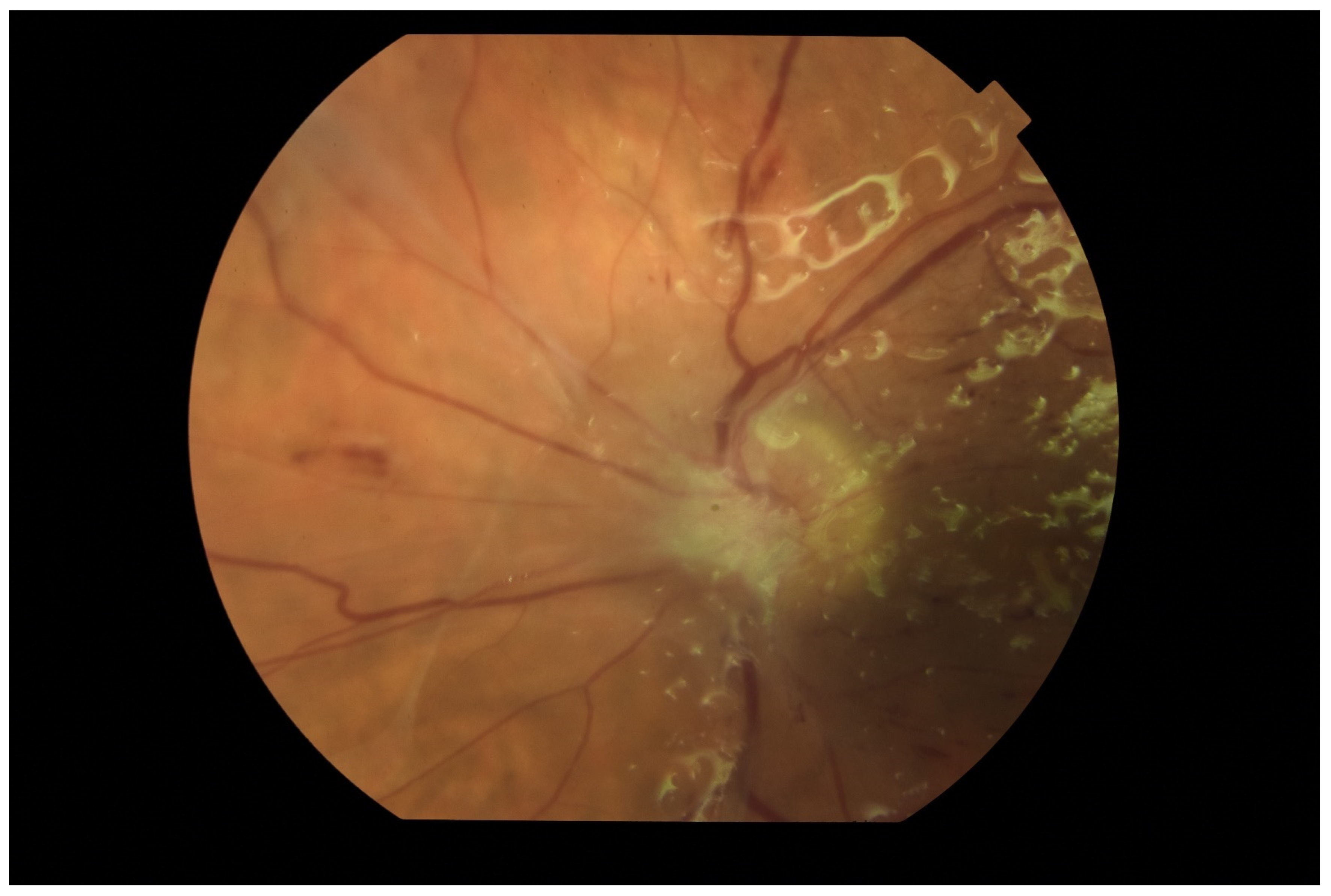
Anterior segment examination revealed mild anterior chamber cells and flare. Fundus examination showed multiple creamy-yellow retinal infiltrates with overlying vitreous haze, obscuring the macula.
Optical coherence tomography (OCT) revealed intraretinal hyperreflective foci and neurosensory detachment, consistent with inflammatory retinal abscesses.
B-scan ultrasonography demonstrated dense vitreous opacities and choroidal thickening. Blood culture grew Klebsiella pneumoniae, indicating a systemic source of infection.
The diagnosis was endogenous endophthalmitis with retinal involvement secondary to a liver abscess.
Intravitreal and systemic antibiotics were initiated, followed by pars plana vitrectomy, leading to partial recovery of retinal structure and vision.
Disease Entity
Endogenous Endophthalmitis (EE) is a retina-threatening intraocular infection resulting from hematogenous dissemination of pathogens from a distant systemic focus.
Unlike exogenous forms, which occur after surgery or trauma, EE develops internally when microorganisms infiltrate ocular tissues through the bloodstream.
The retina and choroid are often the first ocular structures affected due to their rich vascular supply, making the posterior segment the main site of inflammation and tissue damage.
Fundus Explorer Pro
Photograph the retinal findings described here with the phone already in your pocket — 22 D optics and built-in illumination in one handheld unit.
From Choroida — the team behind this sitePathophysiology
The pathogenesis of retinal involvement in EE begins when bacteria or fungi enter the choroidal or retinal circulation via the posterior ciliary or central retinal arteries.
Once lodged in small vessels, these organisms trigger localized vasculitis and microabscess formation within the retina or choroid.
Infection may progress as follows:
-
Choroidal seeding: Pathogens initially settle in the choroidal capillaries, leading to focal choroiditis.
-
Retinal involvement: The infection spreads inward through Bruch’s membrane to the retinal pigment epithelium (RPE) and retina, forming white infiltrates or retinal abscesses.
-
Vitreous extension: Inflammatory debris and organisms enter the vitreous cavity, producing dense vitritis.
-
Secondary retinal complications: These include retinal necrosis, vascular occlusion, or tractional retinal detachment.
The retinal damage results from both direct microbial destruction and host inflammatory responses, leading to scarring and photoreceptor loss if untreated.
Epidemiology
Endogenous endophthalmitis is relatively rare, representing 2–15% of all endophthalmitis cases, but the retinal consequences are often devastating.
-
Pathogen variation:
-
Klebsiella pneumoniae is the leading cause in East Asia, associated with liver abscesses.
-
Staphylococcus aureus, Streptococcus spp., and Candida albicans predominate in Western populations.
-
-
Systemic conditions: Diabetes mellitus, malignancy, and immunosuppression are major risk factors.
-
Laterality: The condition is unilateral in 70–80% of patients, but can be bilateral in systemic fungemia.
Retinal Clinical Features
The retinal manifestations of endogenous endophthalmitis vary depending on the infecting organism and stage of infection.
Early retinal findings:
-
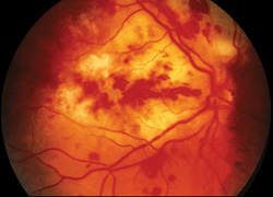
Creamy or yellow-white infiltrates within the retina or subretinal space.
-
Retinal hemorrhages surrounding the lesions.
-
Cotton-wool–like inflammatory foci in Candida infections (“cotton ball” lesions).
Advanced findings:
-
Dense vitritis obscuring retinal details.
-
Retinal abscess formation.
-
Chorioretinal scars after healing.
-
Retinal detachment due to necrosis or tractional forces.
-
Optic disc edema and peripapillary inflammation.
OCT imaging often shows hyperreflective retinal layers, disorganization of the outer retina, and RPE elevation, whereas fluorescein angiography (FA) demonstrates early hypofluorescence (due to blockage) and late leakage from inflamed vessels.
Systemic Associations
The retinal infection originates from systemic foci, commonly including:
-
Liver abscess (Klebsiella pneumoniae)
-
Infective endocarditis
-
Urinary tract infections
-
Infected catheters
-
Pneumonia
-
Soft tissue infections
-
Intravenous drug use (especially fungal causes)
Thus, recognizing retinal findings can serve as an early clue to underlying systemic sepsis, prompting life-saving investigations.
Examination Findings
-
Anterior segment: Mild to moderate anterior chamber reaction, hypopyon in severe cases.
-
Posterior segment:
-
Multifocal retinal infiltrates and hemorrhages.
-
Vitreous haze or snowball-like opacities.
-
Cotton-ball lesions with “string of pearls” appearance in fungal cases.
-
Retinal vascular sheathing and perivascular infiltrates.
-
-
B-scan ultrasonography: Detects vitreous opacities, choroidal thickening, and exudative retinal detachment when fundus view is poor.
-
OCT: Demonstrates retinal abscess cavities, intraretinal hyperreflective foci, and outer retinal disruption.
Differential Diagnosis
-
Bacterial or viral retinitis (e.g., CMV, HSV)
-
Toxoplasma retinochoroiditis
-
Tuberculous choroiditis
-
Ocular lymphoma
-
Acute retinal necrosis
-
Fungal chorioretinitis
Distinguishing features include the presence of systemic infection, positive blood cultures, and bilateral involvement in fungal cases.
Diagnosis
A rapid, multidisciplinary approach is vital.
Ophthalmic evaluation:
-
Fundus photography, OCT, FA, and B-scan imaging to document retinal and vitreous involvement.
Microbiological testing:
-
Blood cultures (positive in 50–70% of cases).
-
Vitreous or aqueous taps for Gram stain, culture, and PCR.
Systemic investigations:
-
Complete blood count, ESR, and CRP.
-
Imaging (CT/MRI/ultrasound) to locate the primary infection.
Management
Early, aggressive treatment is essential to preserve retinal integrity and vision.
1. Systemic Antimicrobial Therapy
-
Broad-spectrum intravenous antibiotics are initiated immediately and adjusted after culture results.
-
Vancomycin + Ceftazidime for bacterial infections.
-
Fluconazole or Voriconazole for fungal cases.
-
-
Duration: 2–4 weeks or until systemic infection is eradicated.
2. Intravitreal Therapy
-
Ensures high intraocular drug concentration:
-
Vancomycin 1 mg/0.1 mL + Ceftazidime 2.25 mg/0.1 mL.
-
Amphotericin B or Voriconazole for fungal infections.
-
3. Pars Plana Vitrectomy (PPV)
-
Indicated in cases with severe vitritis or no improvement after intravitreal therapy.
-
Removes infected vitreous material and inflammatory debris.
-
Reduces microbial load and retinal traction.
4. Retinal Care During Surgery
-
Avoid retinal tears or detachments during vitrectomy.
-
Endolaser photocoagulation around retinal abscesses may be used to limit spread.
5. Source Control
-
Drain abscesses or remove infected catheters.
-
Manage underlying systemic disease such as diabetes or malignancy.
Prognosis
Despite advances in antimicrobial therapy, retinal outcomes remain guarded.
-
Up to 50% of affected eyes end with poor visual acuity (<20/200).
-
Prognosis depends on pathogen virulence, time to treatment, and extent of retinal destruction.
-
Early vitrectomy improves visual outcomes by preventing macular involvement and retinal detachment.
Prevention
-
Prompt treatment of systemic infections.
-
Routine fundus screening for high-risk patients (especially with Klebsiella sepsis or candidemia).
-
Maintaining good glycemic control in diabetics.
-
Aseptic handling of intravenous catheters.


Document what you see
Two smartphone imaging tools built for everyday clinic use — one for the slit lamp, one for the fundus.
From Choroida — the team behind this siteReferences
-
Jackson TL, et al. Endogenous bacterial endophthalmitis: a 17-year prospective series and review of 267 cases. Surv Ophthalmol. 2003;48(4):403–423.
-
Lingappan A, et al. Endogenous fungal endophthalmitis: causative organisms, management strategies, and visual outcomes. Am J Ophthalmol. 2012;153(1):162–166.
-
Tanaka T, et al. Retinal manifestations and OCT features of endogenous endophthalmitis. Ophthalmology Retina. 2018;2(2):140–150.
-
Wong JS, et al. Endogenous bacterial endophthalmitis: an East Asian experience. Ophthalmology. 2000;107(8):1483–1491.
Test yourself
A few questions straight from this article.
-
How does infection reach the eye in endogenous endophthalmitis?
Endogenous endophthalmitis arises when pathogens disseminate through the bloodstream from a distant focus, unlike exogenous forms that follow surgery or trauma. -
What share of all endophthalmitis cases is endogenous in origin?
Endogenous endophthalmitis is relatively rare, accounting for 2-15% of all endophthalmitis, yet its retinal consequences are often devastating. -
Which organism is the leading cause of endogenous endophthalmitis in East Asia, linked to liver abscess?
Klebsiella pneumoniae, typically arising from a liver abscess, is the leading cause in East Asia, while staphylococci, streptococci and Candida predominate in Western populations. -
How often is endogenous endophthalmitis unilateral at presentation?
The condition is unilateral in 70-80% of patients, but bilateral involvement is characteristic of systemic fungemia. -
Which vitreous finding suggests a fungal cause of endogenous endophthalmitis?
Fungal cases classically show cotton-ball inflammatory foci with a string-of-pearls appearance in the vitreous, described particularly with Candida infection. -
What pattern does fluorescein angiography show over retinal lesions in endogenous endophthalmitis?
Fluorescein angiography demonstrates early hypofluorescence caused by blockage from the infiltrates, followed by late leakage from inflamed vessels. -
How often are blood cultures positive in endogenous endophthalmitis?
Blood cultures are positive in 50-70% of cases, and vitreous or aqueous taps for Gram stain, culture and PCR complement them. -
Which intravitreal combination is used for bacterial endogenous endophthalmitis?
Intravitreal vancomycin 1 mg/0.1 mL with ceftazidime 2.25 mg/0.1 mL delivers high intraocular concentrations; amphotericin B or voriconazole is used for fungal cases. -
When is pars plana vitrectomy indicated in endogenous endophthalmitis?
Vitrectomy is indicated for severe vitritis or lack of improvement after intravitreal therapy, and early surgery improves outcomes by limiting macular involvement and detachment. -
What is the visual outcome in endogenous endophthalmitis despite modern antimicrobial therapy?
Up to 50% of affected eyes end with visual acuity below 20/200; outcome depends on pathogen virulence, time to treatment and the extent of retinal destruction.
