
Case Study
A 45-year-old woman presented with restricted eye movements, redness, and discomfort in her right eye following a severe episode of Stevens–Johnson syndrome (SJS) six months earlier.
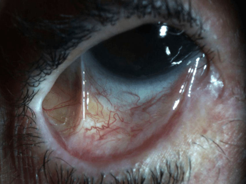
On examination, the lower conjunctival fornix was obliterated, and fibrous adhesions were seen between the bulbar and palpebral conjunctiva.
The patient had reduced tear meniscus height, mild corneal vascularization, and limitation of ocular motility.
Based on these findings, a diagnosis of symblepharon formation secondary to Stevens–Johnson syndrome was made.
Disease Entity
Symblepharon formation refers to the adhesion or scarring between the palpebral (eyelid) and bulbar (eyeball) conjunctiva.
This fibrotic connection restricts ocular motility, causes chronic irritation, and may distort the fornices or eyelid position.
The condition often develops after severe ocular surface inflammation, chemical burns, autoimmune disorders, or trauma.
The extent of adhesion may vary from small localized bands to complete obliteration of the conjunctival fornices, significantly affecting visual function, ocular comfort, and cosmesis.
All-fit Slit-Lamp Adapter
Record and share exactly what you see at the slit lamp. One adapter fits any slit lamp or surgical microscope — and any smartphone.
From Choroida — the team behind this sitePathophysiology
The formation of symblepharon is the result of conjunctival epithelial loss followed by fibroblastic proliferation and scarring.
When the normal conjunctival epithelium is destroyed, the underlying subepithelial tissue becomes exposed and inflamed.
During healing, the raw bulbar and palpebral surfaces adhere to each other, leading to the formation of fibrous bands.
Chronic inflammation, persistent epithelial defects, and repeated trauma perpetuate fibrosis. In severe cases, complete fornix obliteration and ankyloblepharon (fusion of eyelids) can occur.
Causes
Symblepharon may result from various inflammatory, traumatic, or iatrogenic causes, including:
-
Chemical or thermal burns – particularly alkali burns
-
Stevens–Johnson syndrome / Toxic epidermal necrolysis
-
Ocular cicatricial pemphigoid (OCP)
-
Severe conjunctivitis (e.g., membranous or pseudomembranous)
-
Post-surgical scarring (after pterygium or strabismus surgery)
-
Mechanical trauma or radiation injury
-
Infectious causes – such as trachoma
-
Chronic use of topical medications leads to toxic conjunctivitis
Clinical Features
Patients with symblepharon often present with:
-
Restricted ocular motility due to conjunctival adhesions
-
Foreign body sensation, dryness, and redness
-
Decreased fornix depth causing difficulty in eye movement or prosthesis fitting (in anophthalmic patients)
-
Cosmetic deformity or eyelid malposition
-
Corneal complications, such as exposure keratopathy or secondary vascularization
Examination Findings (Summarized):
-
Visible fibrous adhesion between the bulbar and palpebral conjunctiva
-
Fornix shortening or obliteration
-
Limitations of extraocular movements
-
Tear film instability and keratinization ofthe ocular surface
-
Secondary dry eye signs on Schirmer’s test
-
Fluorescein staining showing epithelial irregularity
Classification
Symblepharon can be classified according to extent or severity:
-
Localized – Small adhesion involving a limited conjunctival area.
-
Partial – Involvement of one fornix but sparing ocular motility.
-
Total – Complete fornix obliteration and severe motility restriction.
-
Recurrent – After failed surgical correction due to ongoing inflammation.
Differential Diagnosis
-
Conjunctival inclusion cysts
-
Pterygium recurrence
-
Conjunctival fibrosis secondary to trauma or infection
-
Ocular surface squamous neoplasia (OSSN)
These conditions may mimic adhesions but differ in etiology and treatment approach.

Diagnostic Approach
Diagnosis of symblepharon is largely clinical, based on slit-lamp examination and assessment of ocular motility.
However, ancillary tests are important for evaluating underlying causes and surgical planning:
-
Ocular surface staining with fluorescein or lissamine green to assess epithelial defects.
-
Schirmer’s test and tear breakup time (TBUT) for dry eye evaluation.
-
Conjunctival biopsy for immunofluorescence in suspected OCP.
-
Anterior segment OCT to assess conjunctival architecture and adhesion depth.
Management
Treatment of symblepharon depends on severity, underlying cause, and degree of functional impairment.
The goals are to restore ocular surface integrity, preserve motility, and prevent recurrence.
1. Medical Management
-
Treat the underlying cause (e.g., control inflammation in OCP or SJS).
-
Lubricating drops and ointments to reduce friction.
-
Topical corticosteroids or cyclosporine for inflammatory control.
-
Bandage contact lenses to protect the cornea and minimize adhesion formation.
2. Surgical Management
Surgery is indicated in moderate to severe cases or when vision and motility are affected.
Surgical Techniques Include:
-
Symblepharon lysis: Careful dissection and separation of adhesions.
-
Amniotic membrane transplantation (AMT): Used to reconstruct the conjunctival surface and reduce fibrosis.
-
Conjunctival or mucous membrane grafting: Buccal mucosa or nasal mucosa used to replace lost tissue.
-
Fornix reconstruction: Maintains fornix depth using conformers, symblepharon rings, or scleral shells.
-
Use of antimetabolites (mitomycin C): Prevents recurrence in selected cases.
-
Sutureless techniques with fibrin glue to minimize postoperative scarring.
3. Postoperative Care
-
Regular lubrication and anti-inflammatory drops.
-
Maintenance of ocular surface moisture using punctal plugs or moisture chambers.
-
Frequent follow-up to monitor for recurrence or infection.
Prognosis
The prognosis depends on the cause, extent of fibrosis, and success of surgical reconstruction. Early recognition and intervention yield better outcomes.

Chronic cicatrizing diseases such as OCP or SJS have higher recurrence rates and require lifelong monitoring.
Functional recovery of ocular motility and cosmetic improvement can be achieved in most cases with appropriate surgical planning and postoperative care.
Prevention
Preventing symblepharon formation involves early management of ocular burns, infections, and autoimmune disorders.
-
Immediate irrigation and surface protection after chemical injuries.
-
Use of amniotic membrane grafts in acute ocular burns or SJS to prevent adhesion formation.
-
Early control of inflammation in OCP with systemic immunosuppressive therapy.


Document what you see
Two smartphone imaging tools built for everyday clinic use — one for the slit lamp, one for the fundus.
From Choroida — the team behind this siteReferences
-
Foster CS, et al. Symblepharon Formation: Pathophysiology and Management Strategies. Ophthalmology.
-
Sharma N, et al. Surgical Management of Symblepharon: Techniques and Outcomes. Cornea.
-
Kheirkhah A, et al. Amniotic Membrane Transplantation in Ocular Surface Reconstruction. Br J Ophthalmol.
-
Holland EJ, et al. Ocular Surface Complications of Stevens–Johnson Syndrome. Ophthalmology.
-
Tauber J, et al. Ocular Cicatricial Pemphigoid: Clinical Features and Management. Surv Ophthalmol.
-
Tseng SCG. Role of Amniotic Membrane in Preventing Symblepharon Formation. Cornea.
Test yourself
A few questions straight from this article.
-
What defines symblepharon formation?
Symblepharon is a fibrotic adhesion between eyelid and eyeball conjunctiva that restricts motility, causes chronic irritation and can distort the fornices. -
Which sequence leads to symblepharon formation?
Once the conjunctival epithelium is destroyed, exposed inflamed subepithelial tissue heals by fibroblastic proliferation, and the raw bulbar and palpebral surfaces adhere as fibrous bands. -
Which injury is a classic cause of symblepharon formation?
Chemical and thermal burns, particularly alkali injuries, are classic causes, alongside Stevens-Johnson syndrome, ocular cicatricial pemphigoid and trachoma. -
Which end-stage complication follows severe symblepharon formation?
Continued fibrosis can obliterate the fornix completely and progress to ankyloblepharon, in which the eyelids become fused. -
How is total symblepharon classified in terms of severity?
The classification runs localised, partial, total and recurrent; total denotes complete fornix obliteration with severe restriction of ocular movement. -
Which test is indicated when ocular cicatricial pemphigoid underlies a symblepharon?
Diagnosis is mainly clinical, but conjunctival biopsy with immunofluorescence confirms suspected ocular cicatricial pemphigoid and guides treatment. -
Which symptom typically brings a patient with symblepharon to the clinic?
Adhesions restrict ocular motility and produce foreign body sensation, dryness and redness; shallow fornices also complicate prosthesis fitting in anophthalmic patients. -
Which procedure reconstructs the conjunctival surface after symblepharon lysis?
After adhesions are dissected, amniotic membrane transplantation rebuilds the conjunctival surface and reduces fibrosis; buccal or nasal mucous membrane grafts replace larger tissue defects. -
Which agent is used during symblepharon surgery to prevent recurrence?
The antimetabolite mitomycin C is used in selected cases to limit recurrent fibrosis, while fibrin glue allows sutureless repair with less postoperative scarring. -
Which patients with symblepharon have the highest recurrence risk?
Chronic cicatrising conditions such as ocular cicatricial pemphigoid and Stevens-Johnson syndrome recur more often and need lifelong monitoring.
