
Case Study
A 34-year-old male with no significant medical history presented with complaints of night blindness since childhood, although he had normal vision during the day.
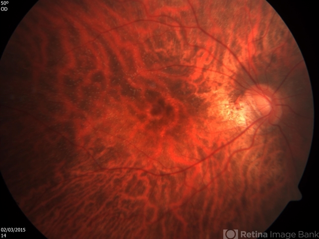
His visual acuity was 20/25 in both eyes, with no significant visual field loss. On funduscopic examination, small, white dots were observed scattered throughout the retinal background.
These dots were most prominent in the macular and mid-peripheral areas. Electroretinography (ERG) confirmed diminished rod function, consistent with a diagnosis of fundus albipunctatus.
Genetic testing revealed a mutation in the RDH5 gene, confirming the diagnosis of this rare condition.
Disease Entity
Fundus albipunctatus (FA) is a rare, inherited retinal disorder characterized by the presence of multiple small, white-yellowish dots scattered throughout the retina.
These dots, which appear as small, discrete lesions, are typically seen in the macula and peripheral retina. The condition is a type of stationary night blindness that affects rod photoreceptors in the retina, leading to a profound impairment in dark adaptation.
The condition is usually non-progressive, and individuals typically have normal central vision during the daytime.
Fundus albipunctatus is a form of congenital stationary night blindness (CSNB) and is most often inherited in an autosomal recessive manner.
It is associated with mutations in the RDH5 gene, which encodes an enzyme involved in retinoid metabolism essential for rod function.
All-fit Slit-Lamp Adapter
Record and share exactly what you see at the slit lamp. One adapter fits any slit lamp or surgical microscope — and any smartphone.
From Choroida — the team behind this sitePathophysiology
The pathophysiology of fundus albipunctatus involves dysfunction of the photoreceptors in the retina, particularly the rods, which are responsible for vision in low-light conditions.
The retinal pigment epithelium (RPE) and rod cells in individuals with FA do not properly process retinoid molecules, which are crucial for the phototransduction cycle.
As a result, rods become less sensitive, impairing the ability to adapt to low-light conditions. This leads to night blindness and a delayed recovery of vision in dim environments.
The white dots observed in the fundus are believed to represent lipofuscin-like deposits within the retina, likely due to the accumulation of incomplete retinoid metabolism products.
These deposits are typically located in the macula and mid-peripheral retina but do not cause significant structural damage to the retina.
Epidemiology
Fundus albipunctatus is considered a rare condition, with estimates suggesting that it accounts for only a small percentage of all cases of congenital stationary night blindness.
- Incidence: It is not fully known how prevalent FA is, but it is considered a rare disorder, with only a few hundred documented cases worldwide.
- Inheritance: The disorder is most commonly inherited in an autosomal recessive manner. In some instances, it can also be inherited in an autosomal dominant pattern, though this is less common.
- Genetic Mutations: The most common mutation associated with FA involves the RDH5 gene. Other mutations, although rare, have been implicated in the condition, leading to varying degrees of clinical presentation.
Clinical Features
The clinical features of fundus albipunctatus are primarily related to visual impairment under low-light conditions.
- Night Blindness: The hallmark symptom of FA is night blindness or difficulty seeing in dim light. This typically presents in childhood, with affected individuals noticing problems with activities such as reading or navigating in poorly lit environments.
- Normal Daytime Vision: In contrast to the difficulties experienced in low-light conditions, patients often maintain normal vision during the day.
- No Significant Visual Field Loss: There is no significant loss of central vision or visual field defects in most cases.
- Funduscopic Findings:
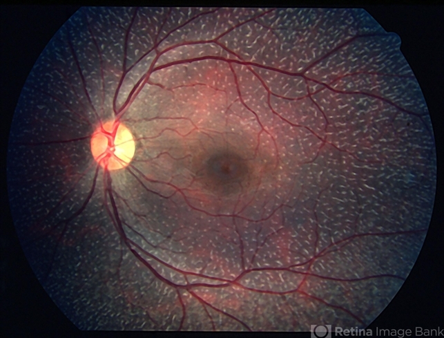
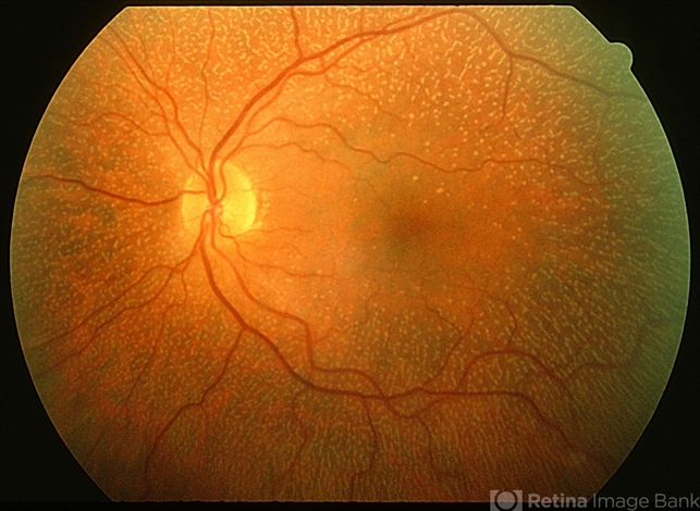
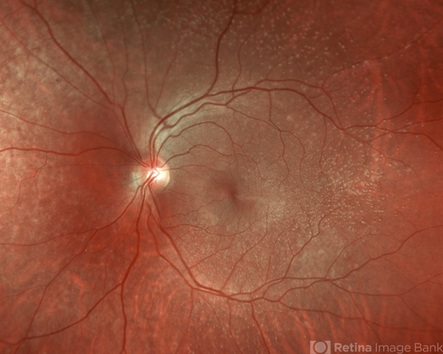
- Numerous, small, white, or yellowish punctate lesions are scattered throughout the retina, most evident in the macula and mid-peripheral retina.
- The white spots appear as discrete, well-defined lesions without significant atrophy or degeneration of the retinal structures.
- Electroretinography (ERG): ERG testing reveals a reduced or absent rod response, indicating impaired rod function. The cone response is typically normal.
Diagnosis
The diagnosis of fundus albipunctatus is based on clinical evaluation, genetic testing, and electroretinography.
- Clinical Examination: The presence of multiple small, white spots in the retina, particularly in the macular and mid-peripheral regions, is characteristic of FA.
- Electroretinography (ERG): ERG testing is essential to confirm the diagnosis, showing a marked reduction in the rod response, while the cone function remains relatively unaffected.
- Genetic Testing: Mutation analysis of the RDH5 gene can confirm the diagnosis. However, not all cases of FA involve RDH5 mutations, so a negative test does not completely rule out the condition.
Differential Diagnosis
Several other retinal disorders may present with similar features to fundus albipunctatus, and differentiation is crucial.
- Stargardt Disease: Stargardt disease presents with macular atrophy and yellowish fundus lesions, but it is associated with progressive central vision loss, unlike the stable visual acuity seen in FA.
- Retinitis Pigmentosa (RP): RP may present with night blindness and retinal degeneration, but it typically leads to progressive peripheral vision loss and is associated with more generalized retinal atrophy.
- Other Forms of Congenital Stationary Night Blindness (CSNB): Different forms of CSNB, such as those due to mutations in the NYX gene, may present with similar night blindness but lack the characteristic white retinal spots.
- Toxoplasmic Retinitis: Can present with retinal lesions, but the clinical course is typically inflammatory, unlike FA, which is non-progressive.
Management
There is no specific treatment for fundus albipunctatus, as it is primarily a condition of impaired night vision with normal visual acuity during the day.
However, certain interventions and lifestyle adjustments can help manage the condition:
- Vision Aids: Low vision aids for night driving or activities in dim environments, such as enhanced lighting or night vision devices, can help improve the quality of life.
- Patient Education: Educating patients about the importance of good lighting and safety precautions in low-light conditions is crucial.
- Genetic Counseling: As FA is inherited in an autosomal recessive pattern, genetic counseling is recommended for families of affected individuals to understand inheritance risks for future generations.
Prognosis
The prognosis for individuals with fundus albipunctatus is generally good, with normal life expectancy and no significant visual impairment during the day.
- Night Blindness: Night blindness often stabilizes after childhood, with most individuals adapting to their condition.
- Visual Acuity: Central visual acuity remains unaffected, and there is no progression to more severe forms of retinal degeneration.
- Retinal Integrity: The retina generally remains intact without significant degeneration, and visual fields remain stable over time.
Prevention
There is no known method to prevent fundus albipunctatus as it is a genetic condition. However, early diagnosis can help mitigate the impact of night blindness by providing patients with appropriate accommodations.
Conclusion
Fundus albipunctatus is a rare, inherited retinal disorder characterized by night blindness and the presence of small, white spots in the retina.
While the condition is non-progressive and central vision is typically preserved, the visual impairment associated with night blindness can significantly impact quality of life.
Early diagnosis, genetic counseling, and adaptation strategies can help individuals with this condition lead fulfilling lives despite their visual challenges.


Document what you see
Two smartphone imaging tools built for everyday clinic use — one for the slit lamp, one for the fundus.
From Choroida — the team behind this siteReferences
- Berson EL. Retinitis pigmentosa and fundus albipunctatus. Archives of Ophthalmology. 1987;105(10):1443-1447.
- Marmor MF, Wolfson Y, Packer S. The genetics of fundus albipunctatus. Archives of Ophthalmology. 1983;101(3):381-384.
- Collin RW, den Hollander AI, Cremers FP. Fundus albipunctatus and related retinopathies: Molecular genetics and clinical features. The British Journal of Ophthalmology. 2011;95(9):1216-1221.
- Sohocki MM, Pericak-Vance MA, Lupski JR. Genetics of fundus albipunctatus: Clinical implications. Retina. 2001;21(6):557-563.
- Gopinath P, Pande M, Kaur I. The role of RDH5 mutations in fundus albipunctatus: A comprehensive review. Journal of Ophthalmology. 2015;2015:728391.
Test yourself
A few questions straight from this article.
-
Which gene mutation is most commonly associated with fundus albipunctatus?
Fundus albipunctatus is most often caused by mutations in RDH5, which encodes an enzyme of retinoid metabolism essential for rod function. -
Fundus albipunctatus is classified as which category of inherited retinal disease?
The article classifies fundus albipunctatus as a form of congenital stationary night blindness, non-progressive and mainly affecting rod photoreceptors. -
What is the usual inheritance pattern of fundus albipunctatus?
Most cases are inherited in an autosomal recessive manner; autosomal dominant inheritance is described but is much less common. -
Where are the white dots of fundus albipunctatus most prominently seen?
The small white or yellowish punctate lesions are scattered through the retina but are most evident in the macular and mid-peripheral regions. -
The white retinal dots in fundus albipunctatus are believed to represent which deposits?
The dots are thought to be lipofuscin-like deposits resulting from accumulation of incomplete retinoid metabolism products, without significant structural retinal damage. -
What is the characteristic electroretinography pattern in fundus albipunctatus?
ERG shows a markedly reduced or absent rod response confirming impaired rod function, while the cone response is typically normal. -
Which feature best distinguishes fundus albipunctatus from Stargardt disease?
Stargardt disease causes macular atrophy with progressive central vision loss, whereas visual acuity in fundus albipunctatus remains stable. -
Which management approach is appropriate for a patient with fundus albipunctatus?
No specific treatment exists; care centres on vision aids for dim environments, patient education on lighting and safety, and genetic counseling for families. -
What is the expected long-term prognosis in fundus albipunctatus?
Central visual acuity remains unaffected, the retina stays largely intact, and there is no progression to more severe retinal degeneration. -
How do other forms of congenital stationary night blindness, such as NYX-related disease, differ from fundus albipunctatus?
Other CSNB forms, such as those from NYX mutations, produce similar night blindness but do not show the characteristic white retinal dots.
