Hypotony maculopathy is vision loss caused by chorioretinal folds and related fundus changes in an eye with abnormally low intraocular pressure (IOP). Most cases follow glaucoma filtration surgery, and most improve once the clinician finds and treats the leak or the underlying cause. This article covers the causes, diagnosis, differential, and management of hypotony maculopathy.
Case report: hypotony maculopathy after an intravitreal injection
A 52-year-old man lost vision in his right eye (OD) two days after an uncomplicated ranibizumab intravitreal injection with a 30G needle. His history included PE, high myopia, and a pars plana vitrectomy in the OD for retinal detachment. He received monthly ranibizumab for choroidal neovascularization secondary to angioid streaks, with 78 previous injections in the OD.
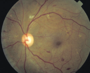
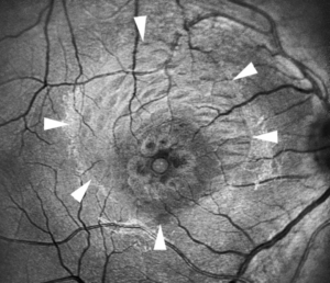
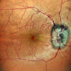
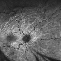
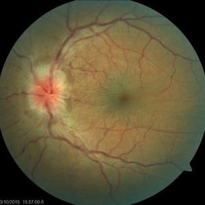
At presentation, best corrected visual acuity (BCVA) was 6/18 and IOP was 3 mmHg. Slit-lamp examination of the anterior segment was unremarkable. Smartphone Fundoscopy and OCT scans revealed posterior pole chorioretinal folds. The low pressure and the fundus findings together established the diagnosis of hypotony maculopathy.
What is ocular hypotony?
Ocular hypotony means abnormally low IOP. Statistically, it is a pressure below 6.5 mmHg, more than 3 standard deviations below the mean. Many eyes at that pressure see normally, so the clinical definition matters more: hypotony is an IOP low enough to cause vision loss.
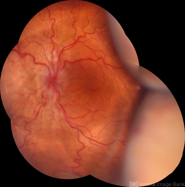
Low pressure reduces vision through corneal edema, astigmatism, cystoid macular edema, or maculopathy. Hypotony maculopathy shows chorioretinal folds and vascular tortuosity, with optic nerve head edema in the acute setting. OCT can reveal folds before they are visible on examination, so image any hypotonous eye with unexplained blur.
Hypotony maculopathy, also called hypotonic maculopathy, follows ocular inflammation, trauma, or surgery. Most cases are secondary to glaucoma filtration surgery, with a reported incidence of 1.3% to 18%. Antifibrosis agents such as mitomycin C raise the risk, as do young age, male sex, and high myopia.
Fundus Explorer Pro
Photograph the retinal findings described here with the phone already in your pocket — 22 D optics and built-in illumination in one handheld unit.
From Choroida — the team behind this siteWhat are the signs and symptoms of hypotony of the eye?
Hypotony of the eye causes blurred vision, distortion, and sometimes a hyperopic shift as folds alter the retinal plane. Examination often shows one or more of the following:
- Shallow anterior chamber or Descemet folds
- Corneal edema
- Choroidal effusion or detachment
- Chorioretinal folds, vascular tortuosity, and disc swelling
Young and highly myopic eyes have lower scleral rigidity, so they develop folds at pressures that older eyes may tolerate.
What causes hypotony?
Hypotony results from reduced aqueous production, increased outflow, or both. Leaks through a wound, bleb, or perforation are the most common mechanism, and ciliary body failure explains many of the rest. The causes of ocular hypotony fall into four groups.
Postoperative hypotony
- Mitomycin C (MMC) toxicity of the ciliary body
- Overfiltration
- Bleb leak
- Wound leak
- Iridocyclitis
- Cyclodialysis
- Ciliochoroidal detachment
- Retinal detachment
Traumatic hypotony
- Scleral perforation
- Retinal detachment
- Cyclodialysis
- Iridocyclitis
- Ciliochoroidal detachment
Bilateral hypotony
- Osmotic dehydration
- Diabetic coma
- Uremia
- Myotonic dystrophy
Miscellaneous forms of hypotony
- Ciliary body hypoperfusion from vascular occlusive disease, such as ocular ischemic syndrome
Intravitreal injections rarely cause hypotony, but a leaking injection site can do so, especially in a thin-walled, highly myopic or vitrectomized eye, which may apply to the eye in our case. Systemic causes such as dehydration and uremia lower pressure in both eyes, while one-sided low pressure should prompt a search for a local leak or detachment.
Hypotony differential diagnosis
When you find low pressure, sort the cause by the anterior segment first. A positive Seidel test points to a wound or bleb leak. A cleft seen on gonioscopy suggests cyclodialysis. A sealed eye with cells and flare points to ciliary body shutdown from inflammation.
Chorioretinal folds also occur at normal pressure, so confirm the IOP before you label the maculopathy. Folds with a normal IOP point elsewhere: choroidal tumor, posterior scleritis, orbital mass, thyroid eye disease, hyperopia, scleral buckling, or papilledema. Disc swelling from hypotony can also mimic raised intracranial pressure.
How do you diagnose hypotony maculopathy?
Diagnosis combines low IOP with characteristic fundus findings. Measure the pressure, perform a Seidel test, inspect the bleb, and dilate the pupil to look for choroidal or retinal detachment. Chorioretinal folds at the macula, tortuous vessels, and disc swelling support the diagnosis.
Tonometry in a very soft eye can be unreliable, so repeat the measurement and compare it with digital palpation. Always check the fellow eye. If both eyes are hypotonous, look for systemic causes such as dehydration, diabetic ketoacidosis, or uremia.
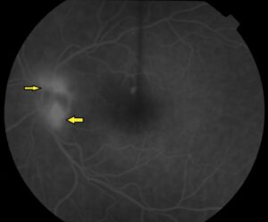
OCT shows folds, retinal thickening, and sometimes subretinal fluid. B-scan ultrasound or ultrasound biomicroscopy shows ciliochoroidal detachment and cyclodialysis clefts. Fluorescein angiography shows alternating hyperfluorescent and hypofluorescent streaks along the folds.
Hypotony maculopathy management
Successful treatment of hypotony maculopathy depends on identifying the underlying cause and treating it. The most common cause is recent glaucoma filtration surgery that produces either a bleb leak or an over-filtering bleb. Trauma and inflammation are other common causes. Match the treatment to the mechanism before you try to raise the pressure.
Medical and conservative management
For a bleb leak, conservative measures include aqueous suppressants, topical antibiotics, and a bandage contact lens. Aqueous suppressants reduce flow through the leak, which may let the overlying epithelium heal. The bandage lens promotes epithelial migration and wound healing, and the antibiotic provides infection prophylaxis. Aminoglycosides may induce conjunctival inflammation that stimulates healing.
If the leak persists, cyanoacrylate glue, autologous blood injection, and compression sutures are further options. For an over-filtering bleb, options include a quick taper of topical steroids, autologous blood injection, laser to stimulate inflammation, diathermy, trichloroacetic acid, and compression sutures that limit aqueous outflow.
When hypotony comes from uveitis and hyposecretion, corticosteroids reduce inflammation and improve ciliary body function, since inflammation lowers aqueous production and increases uveoscleral outflow. Topical cycloplegics relax the iris and ciliary body and reduce the potential space for effusions. Small cyclodialysis clefts may close with cycloplegics or laser, while larger clefts need surgical repair.
Surgical management
Surgery is immediate for trauma, including scleral rupture or retinal detachment, as described in our ruptured globe management strategies article. In other causes, try conservative management first. If these measures fail to resolve the hypotony maculopathy, surgery is warranted.
Bleb excision and revision may be required, using either conjunctival advancement or a free conjunctival autograft. Persistent cyclodialysis clefts can be treated with laser, diathermy, cryopexy, or direct suture repair.
Vitrectomy has been described as a possible treatment for unresolving chorioretinal folds. An internal limiting membrane (ILM) peel may remove contractile forces from subclinical fibrocellular proliferation. Surgeons have also used perfluorocarbon liquid as a steam-roller to flatten the retina, temporarily raising intraoperative IOP to 50 mm Hg.
Hypotony maculopathy prognosis
Vision often recovers when pressure returns to normal and the folds resolve, particularly if you treat the cause early. Long-standing folds can leave permanent damage from retinal pigment epithelium changes and fibrosis, so delay worsens the outlook.
Eyes that stay hypotonous for long periods risk choroidal detachment and, in severe cases, phthisis bulbi. Review any eye with IOP below 6.5 mmHg promptly, repeat OCT after each intervention, and track IOP and acuity until both stabilize.


Document what you see
Two smartphone imaging tools built for everyday clinic use — one for the slit lamp, one for the fundus.
From Choroida — the team behind this siteHow can you prevent hypotony after surgery?
Careful wound construction, tight scleral flap closure, and cautious antimetabolite dosing lower the risk after glaucoma surgery. Warn patients to report blurred vision, and check pressure at early postoperative visits. For eyes that receive repeated injections, such as those treated for age-related macular degeneration, check IOP and the injection site if vision drops afterward.
Test yourself
A few questions straight from this article.
-
What is the statistical definition of ocular hypotony?
Statistically hypotony is an intraocular pressure below 6.5 mmHg, which lies more than three standard deviations below the mean IOP. -
Besides maculopathy, which mechanisms account for vision loss in an eye with very low IOP?
Low IOP can reduce vision through corneal oedema, induced astigmatism, cystoid macular oedema, or maculopathy itself. -
Which fundus findings characterise hypotony maculopathy?
A low IOP combined with chorioretinal folds, acute optic nerve head oedema and vascular tortuosity defines hypotony maculopathy. -
What is the most common cause of hypotony maculopathy?
Most cases follow glaucoma filtration surgery, usually through either a bleb leak or an over-filtering bleb. -
What is the reported incidence of hypotony maculopathy after glaucoma filtration surgery?
Reported incidence after glaucoma filtration surgery ranges from 1.3% to 18%. -
Which intraoperative practice increases the risk of hypotony maculopathy after glaucoma surgery?
The risk of hypotony maculopathy rises when antifibrosis agents are used during glaucoma surgery; mitomycin C toxicity of the ciliary body is one listed postoperative cause. -
How does a bandage contact lens help a leaking bleb causing hypotony?
Conservative measures for a bleb leak include aqueous suppressants, topical antibiotics and a bandage contact lens, which promotes epithelial migration and wound healing. -
Which measure is used to treat an over-filtering bleb causing hypotony?
An over-filtering bleb may be treated with a quick steroid taper, autologous blood injection, laser-induced inflammation, diathermy, trichloroacetic acid or compression sutures. -
In which situation is immediate surgery undertaken for hypotony rather than conservative treatment?
Traumatic cases including scleral rupture or retinal detachment go straight to surgery; for other causes conservative management is tried first and surgery follows only if it fails. -
Which surgical option has been described for unresolving chorioretinal folds in hypotony maculopathy?
Vitrectomy has been described for folds that do not resolve, and an ILM peel may remove contractile forces from subclinical fibrocellular proliferation.

Writes and reviews the clinical articles on Educate. Every post is checked against peer-reviewed sources before it goes live.