CASE REPORT
A 25-year-old male presented to an ophthalmology clinic with a sudden decrease in vision in his left eye. He had a history of keratoconus in both eyes, with the left eye being more affected.
The patient reported waking up with severe eye pain, redness, and a sudden loss of vision in the left eye. On examination, visual acuity in the left eye was counting fingers at 1 meter.
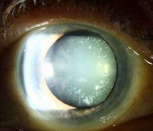
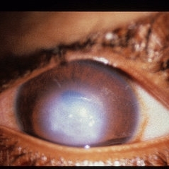
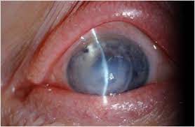
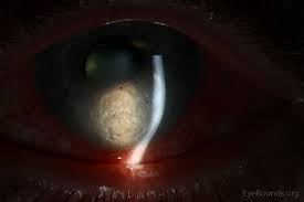
Slit-lamp examination revealed corneal stromal thinning and pronounced edema with Descemet’s membrane folds consistent with acute corneal hydrops. The right eye showed mild corneal steepening but no signs of hydrops.
The patient underwent additional investigations to confirm the diagnosis and rule out other potential causes of corneal edema. Corneal topography was performed, which showed characteristic steepening and irregular astigmatism consistent with advanced keratoconus in both eyes.
Anterior segment optical coherence tomography (AS-OCT) demonstrated corneal thinning, Descemet’s membrane detachment, and the presence of fluid in the corneal stroma, confirming the diagnosis of acute corneal hydrops (ACH).
Acute Corneal Hydrops DISEASE entity
Acute corneal hydrops (ACH), an uncommon complication of corneal ectatic disorders, involves sudden-onset corneal edema due to a rupture in the Descemet membrane (DM) and can cause impaired vision and eye pain.
Acute corneal hydrops (ACH) is believed to result from a break in the Descemet membrane and the endothelium, leading to an influx of aqueous humor into the stroma and subsequent formation of corneal edema. Some histopathologic studies provide evidence that disruption of the posterior stroma may contribute to the pathogenesis as well.
In patients with keratoconus, the incidence of acute corneal hydrops is reported to be between 0.2% and 2.8%. It also occurs rarely in keratoglobus and pellucid marginal degeneration. Acute corneal hydrops presents most commonly between 20 and 40 years of age.
Males may have up to double the risk of females developing ACH. The role of family history in its etiology is highly variable. In New Zealand, individuals of Pacific ethnicity were found to have a higher risk of hydrops compared to individuals of European descent. In the UK, there was a higher prevalence of keratoconus and ACH among South Asian and Black patients.
All-fit Slit-Lamp Adapter
Record and share exactly what you see at the slit lamp. One adapter fits any slit lamp or surgical microscope — and any smartphone.
From Choroida — the team behind this siteMANAGEMENT of Acute Corneal Hydrops
Medical therapy
Initial management of Acute corneal hydrops (ACH)is often conservative, as many cases resolve spontaneously within 2 to 4 months. There is a lack of case-control studies investigating topical treatments of ACH and treatment is largely chosen based on anecdotal evidence.
Topical measures frequently include an ocular antihypertensive, hypertonic saline, cycloplegia, steroid, and an antibiotic. In the presence of Seidel positivity, which may be related to the transudation of aqueous through the edematous cornea instead of perforation, an aqueous suppressant and pressure patching may be utilized.
Surgery
- Air/gas: In cases of ACH where the DM is widely separated from the cornea stroma, a pneumatic descemetopexy may be considered to aid in reattachment. Early intracameral injection of air, C3F8 gas, or SF6 gas has been shown to hasten deturgescence of the cornea in ACH.
- Gas remains in the anterior chamber for a longer duration than air, but requires longer supine positioning with an increased risk of complications. Gases must be used at iso-expansile concentrations. Although both air and gas have been shown to decrease the time until edema resolution, there does not appear to be any improvement in final visual acuity or in the need for later corneal transplantation.
- Compression sutures: Like intracameral air and gas, corneal compression sutures may be used to reapproximate the detached DM. Compression sutures may be particularly useful in the presence of intrastromal clefts. Compression sutures placed perpendicular to the DM tear have been used in isolation or combined with intraoperative air injection.
- Endothelial Keratoplasty: There is recent evidence that endothelial keratoplasty in the acute period after ACH may hasten corneal clearing, improve visual acuity, and lessen the need for later full-thickness transplantation.
- Both Descemet stripping automated endothelial keratoplasty (DSAEK) and Descemet membrane endothelial keratoplasty (DMEK) have been used to restore normal posterior corneal anatomy following an episode of ACH. Long-term follow-up is needed to monitor the formation of corneal scars necessitating more extensive transplantation.
- Penetrating Keratoplasty (PKP)/Deep Anterior Lamellar Keratoplasty (DALK): In cases of resolved ACH that result in a vision-debilitating scar, treatment has traditionally consisted of PKP. While PKP has been shown to have excellent success in keratoconus patients, success rates are reduced in those patients following an episode of ACH, particularly if neovascularization develops.
- Given the long-term complications associated with PKP and the relatively young population of ACH patients, DALK may be considered to remove the scar while preserving the patient’s pre-Descemet layer and DM, although it presents technical challenges in this setting.


Document what you see
Two smartphone imaging tools built for everyday clinic use — one for the slit lamp, one for the fundus.
From Choroida — the team behind this siteREFERENCES
- Barsam A, Petrushkin H, Brennan N, et al. Acute corneal hydrops in keratoconus: a national prospective study of incidence and management. Eye . 2015;29(4):469-474.
- Jump up to:2.0 2.1 2.2 Tuft SJ, Gregory WM, Buckley RJ. Acute corneal hydrops in keratoconus. Ophthalmology. 1994;101(10):1738-1744.
- Grewal S, Laibson PR, Cohen EJ, Rapuano CJ. Acute hydrops in the corneal ectasias: associated factors and outcomes. Trans Am Ophthalmol Soc. 1999;97:187-198; discussion 198-203.
- Sridhar MS, Mahesh S, Bansal AK, Nutheti R, Rao GN. Pellucid marginal corneal degeneration. Ophthalmology. 2004;111(6):1102-1107.
- Jump up to:5.0 5.1 5.2 5.3 5.4 5.5 5.6 Barsam A, Brennan N, Petrushkin H, et al. Case-control study of risk factors for acute corneal hydrops in keratoconus. Br J Ophthalmol. 2017;101(4):499-502.
Test yourself
A few questions straight from this article.
-
Acute corneal hydrops occurs as a complication of which group of disorders?
Acute corneal hydrops is an uncommon complication of the corneal ectatic disorders, producing sudden corneal oedema with impaired vision and eye pain. -
What produces the sudden corneal oedema of acute corneal hydrops?
A break in Descemet membrane and the endothelium allows aqueous humour to flood the stroma; disruption of the posterior stroma may also contribute. -
What is the reported incidence of acute corneal hydrops in patients with keratoconus?
Reported incidence of hydrops among keratoconus patients falls between 0.2 and 2.8 percent. -
Besides keratoconus, acute corneal hydrops occurs rarely in which conditions?
Beyond keratoconus, hydrops is occasionally seen in the other ectatic conditions, keratoglobus and pellucid marginal degeneration. -
At what age does acute corneal hydrops most commonly present?
Acute corneal hydrops presents most commonly between 20 and 40 years of age, and men may carry up to twice the risk of women. -
Which anterior segment OCT findings confirm acute corneal hydrops?
AS-OCT shows corneal thinning, detachment of Descemet membrane and fluid within the corneal stroma, confirming the diagnosis. -
What is the usual initial approach to acute corneal hydrops?
Initial management is usually conservative because many episodes settle spontaneously over two to four months; topical choices rest largely on anecdotal evidence. -
Which topical measures are frequently used in acute corneal hydrops?
Frequent topical measures include an ocular antihypertensive, hypertonic saline, cycloplegia, a steroid and an antibiotic. -
What effect does early intracameral air or gas have in acute corneal hydrops?
Air and gas both hasten deturgescence of the cornea, but neither improves final visual acuity nor reduces the later need for corneal transplantation. -
How does a previous episode of acute corneal hydrops affect penetrating keratoplasty outcomes?
Penetrating keratoplasty does well in keratoconus generally, but success rates are reduced after an episode of hydrops, especially where neovascularisation has developed.

Writes and reviews the clinical articles on Educate. Every post is checked against peer-reviewed sources before it goes live.