
CASE REPORT
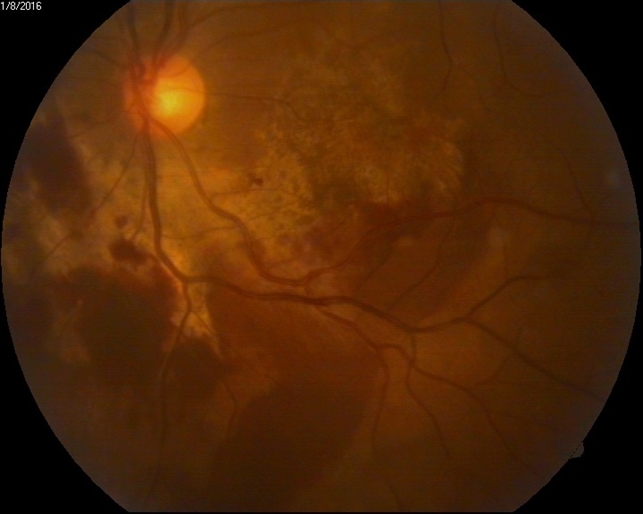
A 12- year- old boy with a history of BB gun injury to his right eye was referred for loss of vision. His visual acuity was counting fingers at one meter in the right eye and with 3+ relative afferent pupillary defect (RAPD).
On slit lamp examination, the right eye appeared normal except for a 1+ vitreous reaction. Fundus examination of the right eye revealed a pale disc with a superior retinal scar and diffuse submacular fibrosis compatible with chorioretinitis sclopetaria.
DISEASE
Chorioretinitis sclopetaria is a closed globe injury causing a full-thickness rupture of the choroid and retina due to blunt trauma from a high-velocity object such as a bullet passing very close to the globe.
Chorioretinal layers split and retract from the sudden strike of the object while the sclera remains intact. Chorioretinitis sclopetaria is a type of coup injury which means the damage is present at the site of impact.
When a high-velocity object passing adjacent to the globe, the direct and indirect shock waves forces cause the chorioretinal layer to rupture followed by vitreous hemorrhage. The retinal pigment epithelium undergoes hyperplasia with extensive loss of photoreceptors.
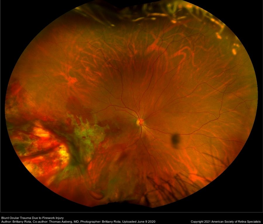
Later on, when the blood absorbs the white fibrous tissue is formed at the site of rupture which is then replaced with dense connective tissues resulting in scarring. The scar has irregular borders like a claw. There is no open globe injury and Intraocular pressure remains within normal limits.
Fundus Explorer Pro
Photograph the retinal findings described here with the phone already in your pocket — 22 D optics and built-in illumination in one handheld unit.
From Choroida — the team behind this siteSigns and symptoms of Chorioretinitis Sclopetaria
The decrease in vision and severity of pain depend upon the site of rupture. The patient either complains of blurry vision or loss of vision.
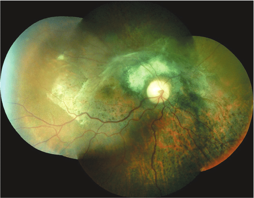
Other findings on dilated fundus examination are areas of bare sclera with adjacent tearing of the retina and choroid, vitreous and subretinal hemorrhages, macular edema, choroidal breaks, and disc edema. After some days fibrous tissues can be seen at the site of chorioretinal hemorrhage on fundus examination when blood is completely reabsorbed.
It is believed that the scar formation at the site of impact would prevent retinal detachment. That’s why in chorioretinitis sclopetaria, the chances of retinal detachment are very low. However, in case of severe orbital injury, the retina could be detached.
Diagnostic procedures
The detailed fundus examination of chorioretinitis sclopetaria can be performed on a slit lamp. However, there are some other diagnostic instruments that can provide further details
- OCT: shows choroidal rupture, the thickness of Retinal pigment epithelium, and retinal detachment.
- B-Scan Ultrasonography: visualizes the vitreous hemorrhage and retinal detachment.
- Fundus Fluorescein Angiography: shows hypofluoresence in the area of chorioretinal rupture.
Chorioretinitis Sclopetaria MANAGEMENT
There is no specific treatment is available for chorioretinitis sclopetaria. The patient should be kept on observation and in case of complications such as retinal detachment, surgery should be performed.
However, this may not improve visual acuity.


Document what you see
Two smartphone imaging tools built for everyday clinic use — one for the slit lamp, one for the fundus.
From Choroida — the team behind this siteREFERENCES
- Sampedro A, Alonso Alvarez C, Ruiz rodriguez M, Usabiaga Bernal JM, Rodriguez vazquez M: Traumatic maculopathies. Arch soc Esp oftalmol. 2001, 76 (1): 57-60.
- Dubovy SR, Guyton DL, Green WR: Clinicopathologic correlation of chorioretinitis sclopetaria. Retina. 1997, 17 (6): 510-20.
- Ludwig CA, Shields RA, Do DV, et al. Traumatic chorioretinitis sclopetaria: Risk factors, management, and prognosis. Am J Ophthalmol Case Rep. 2019;(39-46):39. doi:10.1016/j.ajoc.2019.02.004.
- Shah SU, Prall FR. (2016). Intraorbital Foreign Body Accompanied by Sclopetaria. American Academy of Ophthalmology.
- Dubovy SR, Guyton DL, Green WR. Clinicopathologic correlation of chorioretinitis sclopetaria Retina, 17 (6) (1997), pp. 510-520
- Sadda SR, Wiedemann P, Hinton DR, Schachat AP, Wilkinson CP. Ryan’s Retina. [Electronic Resource]. [London] : Elsevier, 2018.; 2018.
- Blanch R, Ahmed Z, Sik A, et al. (2012). Neuroretinal cell death in a murine model of closed globe injury: pathological and functional characterization.
Test yourself
A few questions straight from this article.
-
What is the defining injury pattern of chorioretinitis sclopetaria?
It is a closed globe injury: the choroid and retina rupture full-thickness and retract, while the sclera remains intact. -
What typically causes chorioretinitis sclopetaria?
Shock-wave forces from a high-velocity object such as a bullet passing very close to the globe rupture the chorioretinal layers. -
What happens to the RPE and photoreceptors in chorioretinitis sclopetaria?
After the rupture and vitreous haemorrhage, the retinal pigment epithelium undergoes hyperplasia with extensive loss of photoreceptors. -
How are the borders of the scar in chorioretinitis sclopetaria described?
As the blood clears, fibrous tissue and then dense connective tissue form a scar whose irregular borders resemble a claw. -
Which dilated fundus finding is characteristic of chorioretinitis sclopetaria?
Fundus findings include areas of bare sclera with adjacent tearing of the retina and choroid, plus vitreous and subretinal haemorrhage. -
Why is retinal detachment uncommon in chorioretinitis sclopetaria?
Scar formation at the impact site is believed to prevent detachment, so the risk is very low unless the orbital injury is severe. -
What does fluorescein angiography show at the rupture site in chorioretinitis sclopetaria?
Fundus fluorescein angiography shows hypofluorescence in the area of the chorioretinal rupture. -
What does B-scan ultrasonography add in the workup of chorioretinitis sclopetaria?
B-scan ultrasonography visualises vitreous haemorrhage and retinal detachment, while OCT shows the choroidal rupture and RPE. -
What is the recommended management of chorioretinitis sclopetaria?
There is no specific treatment; patients are observed, and surgery is reserved for complications such as retinal detachment. -
What is the expected visual effect of surgery for complications of chorioretinitis sclopetaria?
Even when surgery is performed for complications such as retinal detachment, it may not improve visual acuity.
