Reticular pigmentary degeneration is a net-like pattern of pigment in the peripheral fundus of older adults.
It usually lies near or anterior to the equator and is often bilateral. Because the network looks striking on wide-field images, it can worry patients and trainees. However, in most eyes it is a benign aging change that needs observation only.
What Reticular Pigmentary Degeneration Looks Like
The examiner sees fine dark lines forming a polygonal mesh, similar to a fishnet or honeycomb. The cells of the mesh are small and fairly regular, while the retina inside them looks normal, and the pattern often runs circumferentially around the periphery. Retinal vessels cross the network without any change in caliber or course.
Who Develops Reticular Pigmentary Degeneration
The change becomes more common with age and is unusual before late middle age. Autopsy studies described it as a frequent finding in the aging peripheral pigment epithelium. Therefore, its presence in an older patient with good vision should not trigger an extensive workup.
Fundus Explorer Pro
Photograph the retinal findings described here with the phone already in your pocket — 22 D optics and built-in illumination in one handheld unit.
From Choroida — the team behind this siteWhy the Net Pattern Forms
Histopathology links the pattern to aging changes in the retinal pigment epithelium (RPE). Pigment seems to collect along the borders of a polygonal network, while the centers stay relatively pale. However, the exact mechanism is uncertain, so describe it to patients as wear of the pigment layer.
Symptoms in Reticular Pigmentary Degeneration
The change itself causes no symptoms, central acuity is unaffected, and patients also do not notice field loss. As a result, it is almost always an incidental finding on dilated examination or wide-field imaging. If a patient reports night blindness or a narrowing field, look for another cause.
Examination Technique
Indirect ophthalmoscopy with scleral depression shows the network best, and a three-mirror lens also confirms that the retina within the meshes is flat and intact. Check all four quadrants, because the distribution is often patchy, and finally record the extent with a drawing or wide-field photograph for later comparison.
Imaging Reticular Pigmentary Degeneration
Ultra-widefield color imaging captures the whole network in a single view. Fundus autofluorescence may show a mottled or reticular signal, although findings vary between eyes. In addition, peripheral OCT, when feasible, usually shows an intact neurosensory retina with subtle RPE irregularity.
Reticular Pigmentary Degeneration Versus Reticular Pseudodrusen
The shared word “reticular” causes frequent confusion, because reticular pseudodrusen are subretinal drusenoid deposits in the macula and along the arcades. On OCT, they sit above the RPE, and they also carry a recognized risk of late AMD (see reticular pseudodrusen). In contrast, reticular pigmentary degeneration is a peripheral pigment network at the RPE level.
Distinguishing It From Retinitis Pigmentosa
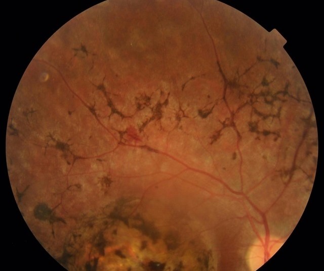
Retinitis pigmentosa produces bone-spicule pigment that often clusters around retinal vessels, and patients also report night blindness, while the disc may show waxy pallor with attenuated arterioles. By comparison, the age-related network is fine and regular, and it spares the disc and vessels. When doubt remains, a markedly reduced electroretinogram points toward a dystrophy.
Other Look-Alikes
Next, consider other causes of peripheral pigment:
- Cobblestone degeneration, with discrete pale lesions and pigmented borders (see cobblestone degeneration)
- Healed chorioretinitis, which leaves irregular atrophic scars
- Drug toxicity, for example thioridazine, with pigment clumping and a relevant drug history
- Old retinal detachment, which may leave pigmented demarcation lines
Link With Macular Degeneration
Some studies report peripheral reticular pigment change more often in eyes with AMD, and an association with complement factor H variants has also been described. However, the finding alone does not predict macular disease in an individual patient. Therefore, examine the macula carefully and manage any AMD on its own features.
Retinal Detachment Risk
Unlike lattice degeneration, this change is not considered a precursor of retinal breaks. It shows no vitreous traction, holes or atrophic thinning on depressed examination. Therefore, prophylactic laser has no role (see peripheral retinal degenerations).
Treatment of Reticular Pigmentary Degeneration
No treatment exists for the network and none is needed, and laser, supplements and topical drops also do not alter it. Instead, treat coexisting disease, such as macular drusen or cataract, on its own merits.
Counseling the Patient
Explain that the pattern reflects normal aging of the pigment layer, and reassure the patient that it does not threaten central vision or cause blindness. Still, teach the usual warning signs of retinal detachment: new floaters, flashes or a curtain.
Follow-Up in Reticular Pigmentary Degeneration
Routine age-appropriate dilated examinations are adequate for most patients, although you should review earlier if new symptoms appear or the macula shows drusen. Compare the periphery with baseline photographs to confirm stability.
Pitfalls to Avoid
- Do not label every peripheral pigment network as retinitis pigmentosa
- Never confuse it with macular reticular pseudodrusen
- Avoid unnecessary laser or repeated imaging
- Do not skip the macular examination in older patients


Document what you see
Two smartphone imaging tools built for everyday clinic use — one for the slit lamp, one for the fundus.
From Choroida — the team behind this siteReferences
- Bastek JV, Siegel EB, Straatsma BR, Foos RY. Chorioretinal juncture. Pigmentary patterns of the peripheral fundus. Ophthalmology. 1982;89:1455-1463.
- Lewis H, Straatsma BR, Foos RY, Lightfoot DO. Reticular degeneration of the pigment epithelium. Ophthalmology. 1985;92:1485-1495.
- Humphrey WT, Carlson RE, Valone JA. Senile reticular pigmentary degeneration. Am J Ophthalmol. 1984;98:717-722.
- Shuler RK Jr, Schmidt S, Gallins P, et al. Peripheral reticular pigmentary change is associated with complement factor H polymorphism (Y402H) in age-related macular degeneration. Arch Ophthalmol. 2008;126:524-527.
- American Academy of Ophthalmology. Basic and Clinical Science Course, Section 12: Retina and Vitreous. San Francisco: AAO; 2023.
Test yourself
A few questions straight from this article.
-
How does reticular pigmentary degeneration typically appear in the peripheral fundus of older adults?
The article describes fine dark lines forming a polygonal mesh, similar to a fishnet or honeycomb, with normal-looking retina inside the cells. -
What does histopathology link reticular pigmentary degeneration to?
Histopathology links the net pattern to aging RPE changes, with pigment collecting along the borders of a polygonal network. The exact mechanism remains uncertain. -
What symptoms does reticular pigmentary degeneration itself typically cause?
The change causes no symptoms, central acuity is unaffected and patients do not notice field loss, so it is almost always found incidentally. -
Which examination method shows the network of reticular pigmentary degeneration best?
Indirect ophthalmoscopy with scleral depression shows the network best. All four quadrants should be checked because the distribution is often patchy. -
How does reticular pigmentary degeneration differ from reticular pseudodrusen?
Reticular pseudodrusen are subretinal drusenoid deposits in the macula and along the arcades, above the RPE, with a risk of late AMD. The pigmentary degeneration is a peripheral RPE-level network. -
Which finding favors retinitis pigmentosa over reticular pigmentary degeneration?
Retinitis pigmentosa causes bone-spicule pigment near vessels, night blindness, waxy disc pallor and attenuated arterioles. The age-related network is fine and regular and spares disc and vessels. -
What test points toward a dystrophy when the diagnosis remains in doubt between a pigment network and retinitis pigmentosa?
When doubt remains, a markedly reduced electroretinogram points toward a dystrophy rather than the benign age-related network. -
Why does reticular pigmentary degeneration not warrant prophylactic laser, unlike lattice degeneration?
Unlike lattice degeneration, it is not considered a precursor of retinal breaks and shows no vitreous traction, holes or atrophic thinning, so prophylactic laser has no role. -
What is the recommended management of reticular pigmentary degeneration?
No treatment exists or is needed; laser, supplements and drops do not alter it. Routine age-appropriate dilated exams are adequate for most patients. -
What does the article advise about the macula in patients with reticular pigmentary degeneration?
Peripheral reticular pigment change is reported more often with AMD, but it does not predict macular disease in an individual. Examine the macula and manage any AMD on its own features.

Writes and reviews the clinical articles on Educate. Every post is checked against peer-reviewed sources before it goes live.