RP vs choroideremia is a frequent question in a patient with night blindness and a shrinking field.
Both cause rod-cone loss, and both can look advanced at the first visit. However, the fundus pattern, family tree and carrier exam usually separate them.
Two Different Diseases
Retinitis pigmentosa (RP) is a group of photoreceptor dystrophies with dozens of causative genes, while choroideremia results from loss of the CHM gene on the X chromosome.
CHM encodes Rab escort protein 1 (REP1), so the RPE, choriocapillaris and photoreceptors degenerate together.
Symptoms in RP vs Choroideremia
Both groups report night blindness first, often in childhood or adolescence, and then the midperipheral field contracts so patients bump into objects.
Central acuity also stays good until late in both diseases, so symptoms alone rarely settle the diagnosis.
Fundus Explorer Pro
Photograph the retinal findings described here with the phone already in your pocket — 22 D optics and built-in illumination in one handheld unit.
From Choroida — the team behind this siteFundus Pattern in RP
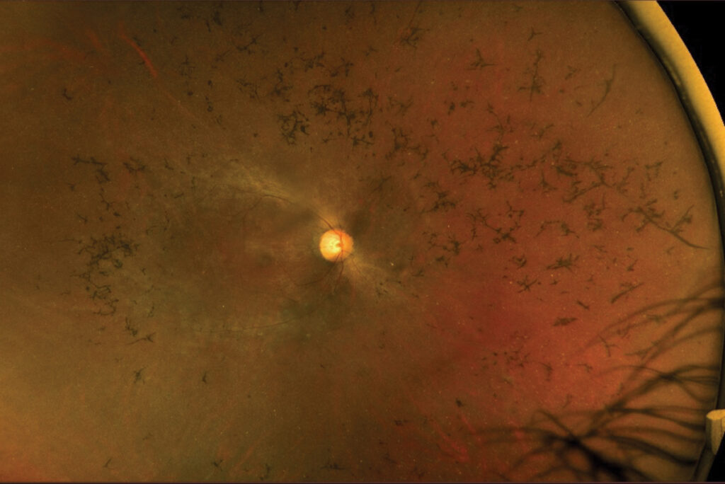
Classic RP shows bone-spicule pigment in the midperiphery, and the arterioles look attenuated while the disc develops waxy pallor.
The triad of spicules, thin vessels and pale disc is the textbook picture. However, early cases may show only subtle RPE mottling.
Fundus Pattern in Choroideremia
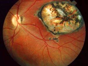
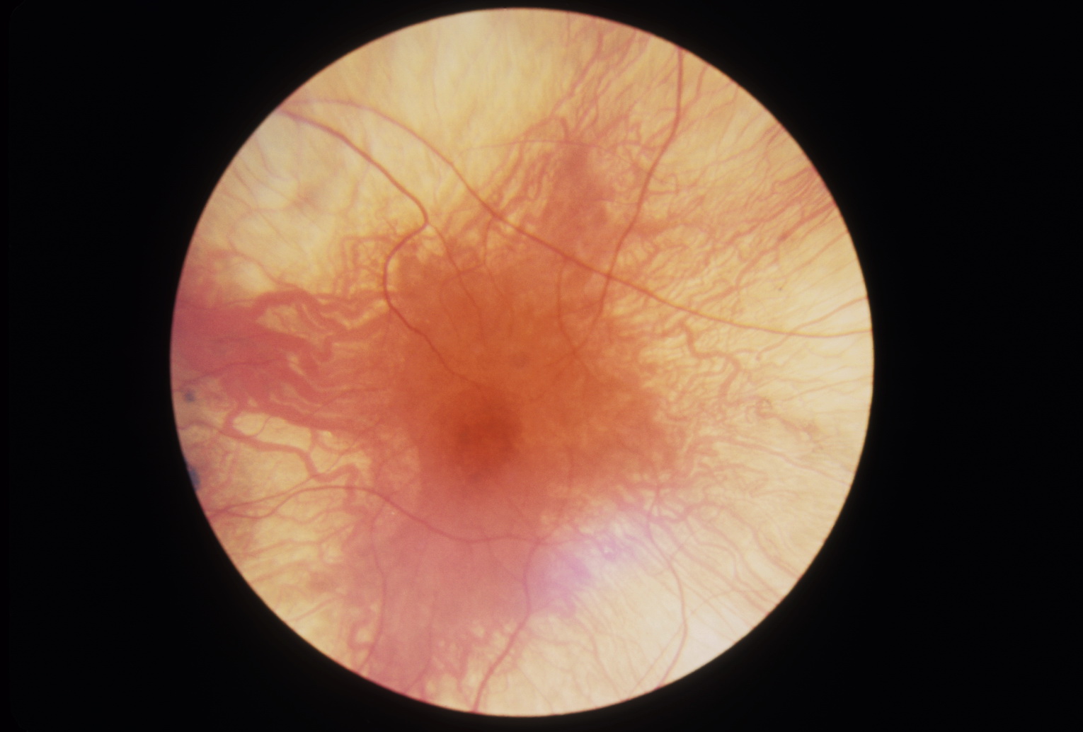
Choroideremia produces scalloped chorioretinal atrophy that starts in the midperiphery, and within these zones, large choroidal vessels and then bare white sclera become visible.
Pigment usually forms fine clumps, while true bone spicules are uncommon, and the vessels and disc often look surprisingly normal until late.
Inheritance in RP vs Choroideremia
RP can follow dominant, recessive or X-linked inheritance, and many cases are simplex, while choroideremia is X-linked recessive, so affected patients are almost always male.
A symptomatic woman with scalloped atrophy may be a manifesting carrier, because of skewed X-inactivation. Therefore, ask about maternal uncles and male cousins on the mother’s side.
Carrier Signs in RP vs Choroideremia
Female choroideremia carriers usually have good vision and no symptoms. However, their periphery often shows patchy, moth-eaten pigment mottling, while carriers of RPGR-related X-linked RP may instead show a tapetal-like reflex at the posterior pole.
For this reason, examine the mother whenever an affected boy presents, because her fundus can then point to the gene before testing returns.
Autofluorescence in RP vs Choroideremia
In RP, autofluorescence often shows a hyperautofluorescent ring around the fovea, which marks the edge of preserved outer retina and shrinks over time.
Choroideremia instead leaves a central island with sharp, scalloped borders (see fundus autofluorescence imaging), and outside it, the atrophic retina looks dark.
OCT in RP vs Choroideremia
OCT in RP shows ellipsoid zone loss that advances from the periphery inward. Cystoid macular edema is common, therefore look for it at every visit.
In choroideremia, OCT shows RPE loss, hypertransmission and a thin choroid, and outer retinal tubulations also line the island border.
Electroretinography
Full-field ERG shows reduced rod responses first in both disorders, and later, cone responses fall, so many adults have extinguished waveforms.
As a result, ERG confirms a rod-cone pattern but cannot separate the two.
Genetic Testing
Panel testing is now standard for both conditions. A pathogenic CHM variant confirms choroideremia, while RP panels must cover many genes.
Absent REP1 protein on blood testing also supports choroideremia when variants are unclear, and then genetic counseling explains risk to siblings, children and female relatives.
Differential Beyond RP vs Choroideremia
- Gyrate atrophy, with high plasma ornithine and scalloped atrophy
- Fundus albipunctatus, with white dots and delayed dark adaptation
- Congenital stationary night blindness, with a nonprogressive course
- Thioridazine toxicity or old inflammatory chorioretinopathy
- Pathologic myopia with extensive chorioretinal atrophy
Gene Therapy Status in RP vs Choroideremia
Regulators have approved voretigene neparvovec for biallelic RPE65 retinal dystrophy, which includes some RP cases (see voretigene neparvovec), but it needs enough viable retinal cells to help.
Several X-linked RPGR programs have also reached late-phase trials with mixed results. For choroideremia, AAV2-REP1 gene therapy showed encouraging early data, but then the phase 3 trial missed its primary endpoint, so no approved treatment exists yet.
Supportive Care and Counseling
First, treat cystoid macular edema in RP with carbonic anhydrase inhibitors, and remove visually significant cataract, while warning about zonular weakness.
Low vision services and mobility training help both groups. Explain that daughters of affected men with choroideremia are obligate carriers.
Finally, a confirmed gene opens trial eligibility (see choroideremia overview).
Pitfalls to Avoid
- Do not label every pigmentary retinopathy as RP without checking the choroid
- Never skip the carrier exam of the mother
- Do not miss treatable macular edema in RP
- Avoid promising gene therapy before a gene is confirmed
- Check ornithine when scalloped atrophy looks atypical


Document what you see
Two smartphone imaging tools built for everyday clinic use — one for the slit lamp, one for the fundus.
From Choroida — the team behind this siteReferences
- Hartong DT, Berson EL, Dryja TP. Retinitis pigmentosa. Lancet. 2006;368:1795-1809.
- Coussa RG, Traboulsi EI. Choroideremia: a review of general findings and pathogenesis. Ophthalmic Genet. 2012;33:57-65.
- Seabra MC, Brown MS, Goldstein JL. Retinal degeneration in choroideremia: deficiency of rab geranylgeranyl transferase. Science. 1993;259:377-381.
- MacLaren RE, Groppe M, Barnard AR, et al. Retinal gene therapy in patients with choroideremia: initial findings from a phase 1/2 clinical trial. Lancet. 2014;383:1129-1137.
- Russell S, Bennett J, Wellman JA, et al. Efficacy and safety of voretigene neparvovec in patients with RPE65-mediated inherited retinal dystrophy: a randomised, controlled, open-label, phase 3 trial. Lancet. 2017;390:849-860.
Test yourself
A few questions straight from this article.
-
Which protein is lost in choroideremia because of CHM gene mutation?
Choroideremia results from loss of the X-linked CHM gene, which encodes REP1, so the RPE, choriocapillaris and photoreceptors degenerate together. -
Which fundus triad is the textbook picture of retinitis pigmentosa?
Classic RP shows midperipheral bone-spicule pigment, attenuated arterioles and waxy disc pallor, though early cases may show only subtle RPE mottling. -
What becomes visible within the atrophic zones of choroideremia as the disease advances?
Within the atrophic zones large choroidal vessels and then bare sclera appear; pigment forms fine clumps and vessels and disc often look normal until late. -
What is the inheritance pattern of choroideremia?
Choroideremia is X-linked recessive, so affected patients are almost always male; a symptomatic woman may be a manifesting carrier from skewed X-inactivation. -
Which peripheral fundus finding is typical of female choroideremia carriers?
Choroideremia carriers usually see well but show patchy moth-eaten peripheral mottling, whereas RPGR-related X-linked RP carriers may show a tapetal-like reflex. -
What does fundus autofluorescence often show in retinitis pigmentosa?
In RP a hyperautofluorescent perifoveal ring marks the edge of preserved outer retina and shrinks over time; choroideremia instead leaves a sharp scalloped central island. -
Which OCT feature lines the border of the preserved central island in choroideremia?
OCT in choroideremia shows RPE loss, hypertransmission and a thin choroid, with outer retinal tubulations lining the island border. -
What can full-field electroretinography establish when comparing retinitis pigmentosa with choroideremia?
ERG shows rod responses reduced first, then cones, in both disorders, so it confirms a rod-cone pattern but cannot separate them. -
Which drug class is used to treat cystoid macular oedema in retinitis pigmentosa?
Cystoid macular oedema is common in RP, should be sought at every visit, and is treated with carbonic anhydrase inhibitors. -
What is the current status of AAV2-REP1 gene therapy for choroideremia?
AAV2-REP1 showed encouraging early data, but its phase 3 trial missed the primary endpoint, so no approved choroideremia treatment exists yet.

Writes and reviews the clinical articles on Educate. Every post is checked against peer-reviewed sources before it goes live.