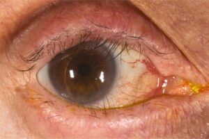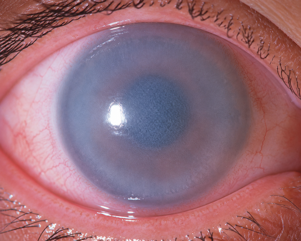
Corneal edema grading turns a vague note like “hazy cornea” into a repeatable record of severity.
The grade combines stromal haze, Descemet folds, epithelial changes and the clarity of the iris view, and pachymetry then adds a number that you can track between visits. As a result, two examiners can judge whether the cornea is recovering.
Why Corneal Edema Grading Helps
Corneal edema changes from day to day after surgery and during keratitis. Therefore, memory alone is a poor guide to progress, and a written grade at each visit shows the direction of change.
How Fluid Enters the Cornea
The endothelium pumps fluid out of the stroma and keeps it relatively dehydrated. When pump function fails, the stroma swells and loses its regular collagen spacing, and light then scatters, so the cornea looks gray.
Raised intraocular pressure can also push fluid into the epithelium, even with a healthy endothelium, and for a structured list of causes, see causes of corneal edema.
All-fit Slit-Lamp Adapter
Record and share exactly what you see at the slit lamp. One adapter fits any slit lamp or surgical microscope — and any smartphone.
From Choroida — the team behind this siteSlit Lamp Technique for Corneal Edema Grading
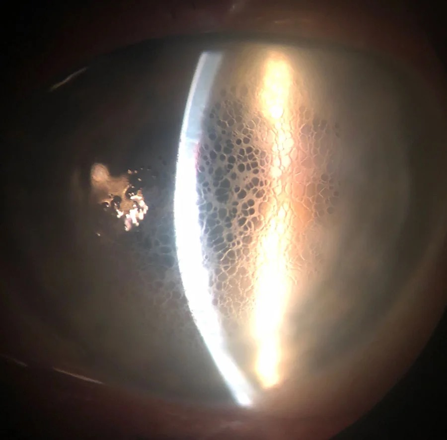
First, use diffuse illumination to judge overall clarity and the iris detail behind the cornea. Next, use a narrow optic section to see stromal thickening and posterior folds. Retroillumination from the fundus reflex shows epithelial microcysts best, while sclerotic scatter reveals subtle central haze.
Stromal Edema Signs
Stromal edema produces a ground-glass haze and a thicker optic section, while Descemet folds appear as fine gray lines in the posterior stroma. Because swelling is greatest where endothelial damage is greatest, the haze often has a visible border.
Epithelial Edema Signs
Epithelial edema appears as fine microcysts or a dull, irregular light reflex, and then microcysts can coalesce into bullae that rupture and cause pain. Patients often report halos around lights and blur that is worse on waking. For example, edema in Fuchs dystrophy often improves through the day as the tear film evaporates.
A Practical Corneal Edema Grading Scale
No single scale has universal acceptance, but many clinics use a simple descriptive scale from 0 to 4.
- Clear (grade 0): normal thickness, no haze, no folds
- Mild (grade 1): faint stromal haze, sharp iris details, few or no folds
- Moderate (grade 2): obvious haze, Descemet folds, slightly blurred iris details
- Marked (grade 3): dense haze, epithelial microcysts, iris details hard to see
- Severe (grade 4): opaque cornea, bullae, no clear view of the iris or pupil
Also record the area involved, for example the central 5 mm or the whole cornea.
Pachymetry and Corneal Edema Grading
Central corneal thickness in healthy adults usually falls in the low to mid 500 micrometer range, although normal values vary widely between individuals. Therefore, a change from the patient’s own baseline tells you more than a single reading. Ultrasound gives a central value, while Scheimpflug imaging maps the whole cornea (see corneal pachymetry).
Subclinical Edema on Tomography
Some corneas look clear at the slit lamp yet already hold excess fluid. Sun and colleagues described Scheimpflug signs of this subclinical edema in Fuchs dystrophy, which include loss of regular isopachs, displacement of the thinnest point and focal posterior surface depression.
Corneal Edema Grading in Fuchs Dystrophy
Krachmer’s classic scale grades guttae by extent, and its highest grade adds stromal or epithelial edema. Later, Repp and colleagues proposed the central-to-peripheral thickness ratio as an objective complement to subjective grading.
Corneal Edema Grading After Cataract Surgery
Mild postoperative edema is common and usually clears within days to weeks (see post-cataract corneal edema). Grade the edema at each visit and record pachymetry when the haze persists. However, edema with raised pressure, a retained nuclear fragment or marked inflammation needs targeted treatment.
Corneal Edema Grading in Keratitis
Edema around an infiltrate reflects inflammation and endothelial stress, so it should shrink as the infection responds. Disciform herpetic keratitis shows edema with keratic precipitates and little infiltrate.
Treatment and Counseling
Hypertonic saline drops or ointment draw fluid from the epithelium and can ease morning blur; however, they do little for deep stromal swelling. Bandage contact lenses can relieve pain from ruptured bullae, and you should explain that persistent grade 3 or 4 edema may need endothelial keratoplasty if the endothelium has failed.
Limits of Grading
Slit lamp grading is subjective and varies between examiners, and scarring, deposits and a poor tear film can mimic or mask edema. In addition, edematous corneas can make applanation pressure readings less reliable.
Pitfalls to Avoid
- Do not grade edema without noting the area involved
- Avoid treating readings from different pachymeters as equivalent
- Remember that morning visits may show worse edema in Fuchs dystrophy
- Check pressure before blaming the endothelium


Document what you see
Two smartphone imaging tools built for everyday clinic use — one for the slit lamp, one for the fundus.
From Choroida — the team behind this siteReferences
- Krachmer JH, Purcell JJ Jr, Young CW, Bucher KD. Corneal endothelial dystrophy: a study of 64 families. Arch Ophthalmol. 1978;96:2036-2039.
- Repp DJ, Hodge DO, Baratz KH, McLaren JW, Patel SV. Fuchs’ endothelial corneal dystrophy: subjective grading versus objective grading based on the central-to-peripheral thickness ratio. Ophthalmology. 2013;120:687-694.
- Sun SY, Wacker K, Baratz KH, Patel SV. Determining subclinical edema in Fuchs endothelial corneal dystrophy: revised classification using Scheimpflug tomography for preoperative assessment. Ophthalmology. 2019;126:195-204.
- Doughty MJ, Zaman ML. Human corneal thickness and its impact on intraocular pressure measures: a review and meta-analysis approach. Surv Ophthalmol. 2000;44:367-408.
- American Academy of Ophthalmology. Basic and Clinical Science Course, Section 8: External Disease and Cornea. San Francisco: AAO; 2024.
Test yourself
A few questions straight from this article.
-
Why does the stroma turn gray when the corneal endothelial pump fails?
The endothelium normally pumps fluid out to keep the stroma relatively dehydrated; when it fails, the stroma swells, loses regular collagen spacing and scatters light. -
Besides endothelial failure, what can drive fluid into the corneal epithelium even when the endothelium is healthy?
Raised pressure can push fluid into the epithelium despite a healthy endothelium, which is why pressure should be checked before blaming the endothelium. -
Which slit lamp illumination shows corneal epithelial microcysts best?
Diffuse light judges overall clarity, an optic section shows stromal thickening and folds, retroillumination shows microcysts and sclerotic scatter reveals subtle central haze. -
How do Descemet folds appear at the slit lamp in stromal corneal edema?
Stromal edema thickens the optic section and produces Descemet folds as fine gray posterior lines, often with a visible border where endothelial damage is greatest. -
In Fuchs dystrophy, how does epithelial edema typically vary through the day?
Patients report halos and blur worse on waking, and Fuchs edema often improves through the day, so morning visits may show worse edema. -
On the 0-4 descriptive corneal edema scale, which grade has dense haze, epithelial microcysts and iris details hard to see?
Grade 2 has obvious haze, folds and slightly blurred iris detail, grade 3 adds dense haze and microcysts, and grade 4 is opaque with bullae. -
Why is a change from baseline pachymetry more useful than a single corneal thickness reading?
Healthy central thickness usually sits in the low to mid 500 micrometer range but varies widely, so a change from the patient's own baseline is more informative. -
Which Scheimpflug finding signals subclinical edema in a Fuchs cornea that looks clear at the slit lamp?
Sun and colleagues described loss of regular isopachs, displacement of the thinnest point and focal posterior surface depression as signs of subclinical edema. -
What is the main limitation of hypertonic saline drops in corneal edema?
Hypertonic saline draws fluid from the epithelium and can ease morning blur, but it has little effect on deep stromal swelling. -
Which corneal edema may need endothelial keratoplasty?
Mild postoperative edema usually clears in days to weeks, but persistent grade 3 or 4 edema may need endothelial keratoplasty if the endothelium has failed.

Writes and reviews the clinical articles on Educate. Every post is checked against peer-reviewed sources before it goes live.