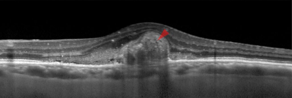
Subretinal hyperreflective material, or SHRM, is a bright layer of tissue between the neurosensory retina and the retinal pigment epithelium on OCT.
It appears in many eyes with neovascular AMD, and it can represent vessels, fibrin, blood, lipid, or scar. Its presence and persistence affect prognosis, so it deserves careful attention.
What Subretinal Hyperreflective Material Is
SHRM is a descriptive term, not a diagnosis, and it includes several tissue types that look similar on OCT: neovascular complexes, fibrin, hemorrhage, vitelliform deposits, and fibrotic scar.
Because the content differs, the response to treatment differs as well. Clinicians use other imaging to separate the types (see macular neovascularization types).
Subretinal Hyperreflective Material in CATT
The Comparison of AMD Treatments Trials analyzed SHRM in detail. Willoughby and colleagues found that SHRM was common at baseline, and it correlated with worse vision at baseline and at two years.
Eyes with SHRM at baseline had a higher risk of scar formation, and its presence at follow-up predicted worse acuity. Jaffe and colleagues reported similar patterns in year five of the trial.
Fundus Explorer Pro
Photograph the retinal findings described here with the phone already in your pocket — 22 D optics and built-in illumination in one handheld unit.
From Choroida — the team behind this siteDifferential Diagnosis of Subretinal Hyperreflective Material
- Active neovascularization with fibrovascular tissue
- Subretinal fibrin from exudation or hemorrhage
- Acute vitelliform material in pattern dystrophies
- Subretinal scar after long-standing disease
Use OCT angiography, fluorescein angiography, and color photographs to identify the source (see submacular hemorrhage management).
Management of Subretinal Hyperreflective Material
Continue anti-VEGF therapy when SHRM reflects active neovascular tissue or exudation. Fibrin and hemorrhage may clear with treatment, and in some eyes the material shrinks over months.
When the material is a scar, additional injections offer little benefit, and visual aids may help. Combination therapy and surgery have a role in selected eyes with thick submacular blood (see anti-VEGF therapy).
Prognosis of Subretinal Hyperreflective Material
The size and the thickness of the material matter, and a thick lesion beneath the fovea carries a worse outlook. Early and sustained treatment lowers the risk of fibrosis, though it cannot prevent it in every eye. Document the presence of SHRM at baseline, and follow its changes with each visit.
Practical Points
- Compare current scans with the baseline instead of only the last visit
- Correlate OCT findings with angiography
- Explain to patients that some material may remain after treatment
An Example Case
A 77-year-old woman starts anti-VEGF therapy for neovascular AMD with acuity of 20/100. OCT shows a thick layer of subretinal hyperreflective material and a small pigment epithelial detachment.
After six monthly injections, the fluid resolves and the material becomes thinner, with a stable border, and vision improves to 20/60, though a patch of material persists. Follow-up shows a fibrotic scar that stays stable on a longer interval.
Questions Patients Ask
Patients ask whether the material is scar tissue, and the answer is that it may be a mix of blood, fibrin, and tissue. They also ask whether injections will remove it, and some material shrinks while scar remains. Because outcomes differ, explain the plan and the goal of keeping vision stable.
Pitfalls to Avoid
- Calling every hyperreflective area a scar
- Stopping treatment because of persistent material without checking for fluid
- Skipping angiography when the diagnosis is uncertain
- Overlooking submacular blood that needs urgent care
Looking Ahead
Artificial intelligence may separate fibrin, vessels, and scar automatically, and new drugs may reduce fibrosis. Until then, follow the material with careful OCT reading.
Practical Points
Compare the width and the height of the material at each visit. Then note changes in the retinal pigment epithelium and the outer retina, and record whether fluid accompanies the material.
Choosing Imaging for Subretinal Hyperreflective Material
OCT angiography shows flow within the material when vessels are present, while fluorescein angiography shows leakage and staining. Because both tests give different information, choose by the clinical question, and also use autofluorescence to detect blood and fibrin, which may look different from scar.
Documenting Subretinal Hyperreflective Material
Record the height, the width, and the location relative to the fovea, then compare the dimensions over time. Finally, note whether the material is increasing, stable, or shrinking. Explain that scars may keep vision low, even when the macula stays dry.
Communicating With Patients
Show the OCT and point out the area of material, then explain that treatment aims to keep the retina stable. Because many patients expect a full recovery, describe realistic goals and remind them to report new distortion promptly.


Document what you see
Two smartphone imaging tools built for everyday clinic use — one for the slit lamp, one for the fundus.
From Choroida — the team behind this siteReferences
- Willoughby AS, Ying GS, Toth CA, et al. Subretinal hyperreflective material in the Comparison of Age-Related Macular Degeneration Treatments Trials. Ophthalmology. 2015;122:1846-1853.
- Daniel E, Toth CA, Grunwald JE, et al. Risk of scar in the Comparison of Age-related Macular Degeneration Treatments Trials. Ophthalmology. 2014;121:656-666.
- Jaffe GJ, Ying GS, Toth CA, et al. Macular morphology and visual acuity in year five of the Comparison of Age-related Macular Degeneration Treatments Trials. Ophthalmology. 2019;126:252-260.
- American Academy of Ophthalmology. Basic and Clinical Science Course, Section 12: Retina and Vitreous. AAO; current edition.
Test yourself
A few questions straight from this article.
-
On OCT, where does subretinal hyperreflective material (SHRM) lie?
SHRM is a bright layer of tissue lying between the neurosensory retina and the retinal pigment epithelium on OCT. -
How is the term subretinal hyperreflective material (SHRM) best classified in neovascular AMD?
SHRM is descriptive, not a diagnosis: neovascular complexes, fibrin, hemorrhage, vitelliform deposits and fibrotic scar can all look alike on OCT, and they respond differently to treatment. -
In the CATT analysis of neovascular AMD, baseline SHRM was associated with a higher risk of which outcome?
In the Comparison of AMD Treatments Trials, eyes with SHRM at baseline had a higher risk of scar and worse vision at baseline and at two years. -
When SHRM in neovascular AMD reflects active neovascular tissue or exudation, what is the recommended management?
Anti-VEGF therapy is continued when SHRM represents active neovascular tissue or exudation; fibrin and blood may clear and the material can shrink over months. -
When SHRM in neovascular AMD represents established scar, what do additional anti-VEGF injections offer?
Scar does not respond the way active tissue does, so further injections offer little benefit and low-vision aids may help instead. -
Which SHRM characteristic carries the worse visual outlook in neovascular AMD?
Size and thickness of the material matter, and a thick lesion beneath the fovea carries a worse outlook. -
Which imaging test shows flow within subretinal hyperreflective material when vessels are present?
OCT angiography shows flow inside the material when vessels are present, whereas fluorescein angiography shows leakage and staining; the test is chosen by the clinical question. -
Which is a recognised pitfall when SHRM persists during anti-VEGF treatment for neovascular AMD?
Persistent material alone is not a reason to stop; fluid must be checked, and not every hyperreflective area should be called scar. -
Which material seen in pattern dystrophies can appear as subretinal hyperreflective material on OCT?
The differential of SHRM includes acute vitelliform material in pattern dystrophies, alongside fibrovascular tissue, fibrin and scar. -
When following subretinal hyperreflective material over time, which scan should the current OCT be compared against?
Current scans should be compared with baseline rather than only the last visit, recording height, width and location relative to the fovea.

Writes and reviews the clinical articles on Educate. Every post is checked against peer-reviewed sources before it goes live.