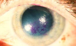
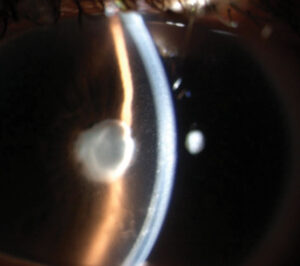
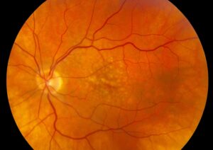
Takayasu retinopathy is the retinal change that occurs in patients with Takayasu arteritis, a large vessel vasculitis that involves the aorta and its branches.
Narrowing of the carotid and subclavian arteries reduces blood flow to the eye.
The retina shows a progressive pattern of ischemic changes that reflects the disease stage.
What Takayasu Retinopathy Is
Takayasu arteritis mainly affects young women, and it causes stenosis of aortic branches.
When the carotid arteries are involved, the eye experiences chronic hypoperfusion.
The fundus then shows dilated capillaries, microaneurysms, and arteriovenous anastomoses (see ocular ischemic syndrome).
Stages of Takayasu Retinopathy
Uyama and Asayama described four stages.
- Dilatation of small vessels marks stage 1
- Microaneurysm formation marks stage 2
- Arteriovenous anastomoses mark stage 3
- Ocular complications such as cataract, rubeosis, and iris atrophy mark stage 4
The stages track the duration and severity of ischemia, though many patients do not show all of them.
Fundus Explorer Pro
Photograph the retinal findings described here with the phone already in your pocket — 22 D optics and built-in illumination in one handheld unit.
From Choroida — the team behind this siteSymptoms of Takayasu Retinopathy
Patients may report amaurosis fugax, blurred vision, and visual loss on standing.
Peter and colleagues studied ocular manifestations in a series and found that retinopathy was frequent.
Some patients have no ocular symptoms but show changes on examination.
Associated Findings in Takayasu Retinopathy
- Pulse deficits and blood pressure differences between arms
- Bruits over the carotid or subclavian arteries
- Headache, fatigue, and systemic inflammation
- Hypertensive retinopathy from renal artery stenosis
Combine findings in the eye with the systemic examination (see hypertensive retinopathy).
Diagnosis of Takayasu Retinopathy
Order inflammatory markers, and refer for vascular imaging with MRA, CTA, or PET.
Fluorescein angiography shows delayed filling, areas of nonperfusion, and leakage at neovascular sites.
Exclude other causes of ischemia, such as giant cell arteritis and atherosclerosis in older patients.
Management of Takayasu Retinopathy
The rheumatology team treats with corticosteroids and immunosuppressants, and sometimes vascular surgery or stenting.
The ophthalmologist monitors for neovascularization and treats with laser or anti-VEGF when needed.
Control of blood pressure protects the retina and the optic nerve.
An Example Case
A 24-year-old woman complains of dimming of vision when she stands up quickly.
Her radial pulses are weak, and her blood pressure differs between arms.
The fundus shows dilated capillaries and a few microaneurysms around the disc, with sluggish filling on angiography.
Imaging of the aorta shows narrowing of the carotid arteries, and she starts immunosuppressive treatment.
Her retinal changes stabilize over a year.
Questions Patients Ask
Patients ask whether the eye changes are reversible, and early stages may improve with control of the disease.
They also ask whether they will lose vision, and the risk depends on ischemia and complications.
Because the disease is chronic, explain the need for long-term follow-up.
Pitfalls to Avoid
- Attributing the retinal findings to diabetes or hypertension alone
- Missing weak pulses in a young woman with visual symptoms
- Delaying referral for vascular imaging
- Treating retinal changes without addressing systemic inflammation
Looking Ahead
Biologic drugs such as tocilizumab may improve control of large vessel vasculitis.
Also, OCT angiography may monitor retinal perfusion without dye.
Therefore, ocular monitoring could become part of disease assessment.
Practical Points
Check blood pressure in both arms and palpate pulses in every young patient with ischemic retinal signs.
Then arrange rheumatology and vascular imaging urgently.
Also screen for neovascular glaucoma and cataract.
Differential Diagnosis
Giant cell arteritis affects older patients, and Takayasu arteritis affects younger women.
Atherosclerotic carotid disease and fibromuscular dysplasia may cause similar retinal findings.
Because the treatment differs, define the cause with imaging.
Surgery and the Eye
Vascular surgery or stenting may restore blood flow, but it may cause a sudden rise in perfusion.
Therefore, monitor the retina for hemorrhages after revascularization.
Also coordinate timing with the ophthalmologist.
Fluorescein Findings in Detail
Angiography shows prolonged arm-to-retina time and slow arterial filling.
Also, it shows capillary dilatation, microaneurysms, and areas of nonperfusion at the periphery.
Because these changes mirror the stage, document them with photographs.
Differential Diagnosis in the Young
Diabetic retinopathy, retinal vein occlusion, and ocular ischemic syndrome may look similar.
However, a young woman with pulse deficits points toward Takayasu arteritis.
Then confirm the diagnosis with vascular imaging, not the eye examination alone.
Pregnancy and Takayasu Arteritis
Pregnancy needs careful planning, since hypertension and flares may occur.
Therefore, coordinate care among obstetrics, rheumatology, and ophthalmology.
Summary
Think of Takayasu arteritis in a young woman with ischemic retinal changes.
Then check pulses and refer for vascular imaging.
Because early treatment protects vessels, do not delay referral.
For the latest studies, see the PubMed results for Takayasu retinopathy.


Document what you see
Two smartphone imaging tools built for everyday clinic use — one for the slit lamp, one for the fundus.
From Choroida — the team behind this siteReferences
- Uyama M, Asayama K. Retinal vascular changes in Takayasu’s disease (pulseless disease), occurring in the early and in the late stages. Doc Ophthalmol Proc Ser. 1976;9:549-554.
- Peter J, David S, Danda D, et al. Ocular manifestations of Takayasu arteritis: a cross-sectional study. Retina. 2011;31:1170-1178.
- Salmon JF. Kanski’s Clinical Ophthalmology: A Systematic Approach. 9th ed. Elsevier; 2020.
- American Academy of Ophthalmology. Basic and Clinical Science Course, Section 12: Retina and Vitreous. AAO; current edition.

Writes and reviews the clinical articles on Educate. Every post is checked against peer-reviewed sources before it goes live.