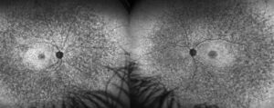
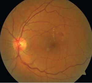
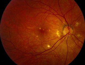
Watershed zones are border regions where two arterial territories meet.
They receive the lowest perfusion pressure in the tissue.
Therefore, they are the first to fail when perfusion falls.
The Concept of Watershed Zones
Each artery supplies a set territory with little overlap.
Where two territories meet, the pressure is lowest.
Also, a drop in systemic or ocular perfusion pressure produces ischemia there first.
As a result, infarcts follow a predictable pattern.
The Choroidal Blood Supply
The choroid receives blood from the short posterior ciliary arteries.
Each artery feeds a wedge-shaped segment of the choroid.
Also, the choriocapillaris lobules are end-arterial units.
Therefore, the choroid contains natural watershed lines between segments.
Fundus Explorer Pro
Photograph the retinal findings described here with the phone already in your pocket — 22 D optics and built-in illumination in one handheld unit.
From Choroida — the team behind this siteHayreh’s Work on Segmental Supply
Hayreh showed that the choroidal circulation is segmental.
He used fluorescein angiography and injections in animals and patients.
Also, he mapped delayed filling at the borders of the segments.
In addition, this work explains many patterns of choroidal ischemia.
Where the Watershed Zones Lie
A key choroidal watershed zone runs near the optic disc and the macula.
It sits between the temporal and nasal short posterior ciliary arteries.
Also, the peripapillary choroid shows the same border effect.
As a result, disc and macular perfusion depend on this zone.
Watershed Zones and Hypotension
A fall in blood pressure lowers flow first in the border zones.
Patients may develop ischemic optic neuropathy after surgery with severe hypotension.
Also, nocturnal dips in pressure add risk in some eyes.
Therefore, blood pressure control must avoid overshoot.
Watershed Zones in Hypertensive Choroidopathy
Malignant hypertension causes fibrinoid necrosis in choroidal vessels.
Focal choriocapillaris infarcts produce Elschnig spots (see hypertensive choroidopathy).
Also, Siegrist streaks mark old linear lesions.
As a result, the pattern follows the lobular arrangement.
Giant Cell Arteritis
Inflammation of the posterior ciliary arteries cuts flow to large choroidal segments.
The disc and the choroid can both show delayed filling.
Also, arteritic ischemic optic neuropathy shows pallid disc swelling.
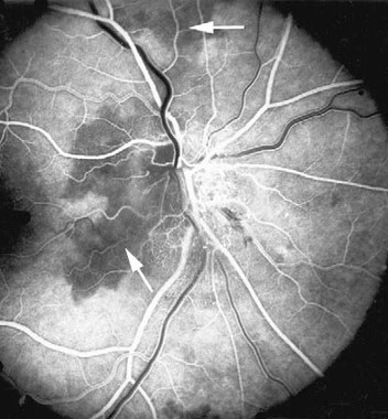
Therefore, fluorescein angiography can show a patchy choroidal filling defect.
Carotid Disease and Ocular Ischemia
Severe carotid stenosis lowers the perfusion of the whole globe.
The retina shows dilated veins and midperipheral hemorrhages.
Also, the choroid shows delayed filling in watershed regions.
In addition, neovascular glaucoma can follow in advanced cases.
Imaging of Watershed Zones
Fluorescein angiography shows delayed filling at the borders in the early frames.
Indocyanine green angiography shows the larger choroidal vessels and their segments.
Also, OCT angiography can show flow voids in the capillary layer.
Therefore, combine modalities for a full map.
Retinal Watershed Areas
The retinal vessels also have border zones in the far periphery and at the foveal edge.
Peripheral nonperfusion first appears there in many diseases.
Also, the foveal avascular zone is a watershed in a sense, since oxygen comes from the choroid.
As a result, the central retina relies on the choroid more than the inner retina.
Optic Nerve Head and Watershed Zones
The optic nerve head receives blood from the short posterior ciliary arteries through the circle of Zinn-Haller.
Border zones around the disc explain the segmental defects in ischemic optic neuropathy.
Also, small crowded discs add vulnerability.
Therefore, the pattern of visual field loss follows the vascular map.
Clinical Implications
Think of the watershed map when a patient has sudden segmental vision loss.
Check blood pressure and consider recent surgery.
Also, look for signs of inflammatory disease.
In addition, consider carotid imaging.
Management Principles
Treatment targets the cause: restore perfusion, treat inflammation and avoid hypotension.
Steroids are urgent for giant cell arteritis.
Also, correct hypovolemia in postoperative cases.
Therefore, early recognition changes outcomes.
Pitfalls to Avoid
- Do not overlook hypotension as a cause of unilateral vision loss
- Never skip inflammatory markers in an older patient with disc swelling
- Do not assume that a normal retinal view excludes choroidal ischemia
- Never ignore delayed choroidal filling on angiography
Ocular Hypoperfusion Syndromes
Low perfusion states include carotid stenosis, aortic arch disease and severe heart failure.
Symptoms can include transient vision loss in bright light.
Also, the retina can show midperipheral hemorrhages.
Therefore, check the carotids and the heart.
Nocturnal Hypotension
Night-time dips in blood pressure can drop flow below the safe level.
Antihypertensive drugs taken at bedtime may add to the dip.
Also, sleep apnea can interact.
As a result, review the timing of medicines.
Surgery and Anemia
Major surgery with blood loss and hypotension can cause ischemic optic neuropathy.
Anemia adds to the risk.
Also, prone spine surgery carries extra risk.
Therefore, monitor pressure and hemoglobin in long cases.
For the latest studies, see the PubMed results for watershed zones.


Document what you see
Two smartphone imaging tools built for everyday clinic use — one for the slit lamp, one for the fundus.
From Choroida — the team behind this siteReferences
- Hayreh SS. Segmental nature of the choroidal vasculature. Br J Ophthalmol. 1975;59:631-648.
- Hayreh SS. Posterior ciliary artery circulation in health and disease: the Weisenfeld lecture. Invest Ophthalmol Vis Sci. 2004;45:749-757.
- Hayreh SS. The blood supply of the optic nerve head and the evaluation of it: myth and reality. Prog Retin Eye Res. 2001;20:563-593.
- Nickla DL, Wallman J. The multifunctional choroid. Prog Retin Eye Res. 2010;29:144-168.

Writes and reviews the clinical articles on Educate. Every post is checked against peer-reviewed sources before it goes live.