Pneumocystis choroidopathy is a choroidal infection that signals disseminated Pneumocystis jirovecii disease in a person with advanced HIV.
Freeman and colleagues first described it in 1989, and it was linked to aerosolized pentamidine prophylaxis.
Today it is rare, but it still occurs in people with undiagnosed HIV or poor access to care.
Clinical Features of Pneumocystis Choroidopathy
Patients usually have few visual symptoms, and the lesions are found on routine dilated examination.
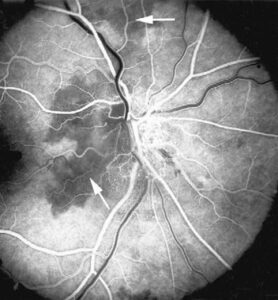
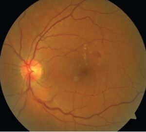
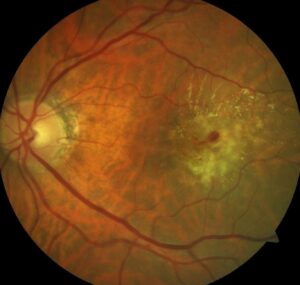
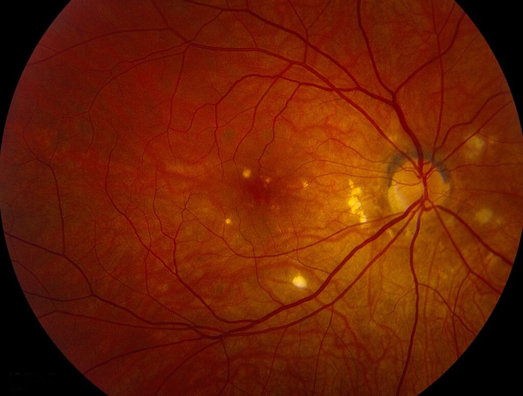
The lesions are creamy, yellow-orange, round or oval patches in the choroid, and they cluster at the posterior pole.
Each patch measures about a quarter to two disc diameters.
Also, the vitreous is quiet, and the overlying retina is usually clear (see HIV retinopathy).
Why Pneumocystis Choroidopathy Occurs
Aerosolized pentamidine protects the lungs but does not reach extrapulmonary sites.
As a result, the organism can spread through the blood to the choroid, the spleen, and the liver.
Rao and colleagues showed foamy eosinophilic infiltrates with organisms in the choroid on histology.
Therefore, the finding means systemic disease, not isolated eye disease.
Fundus Explorer Pro
Photograph the retinal findings described here with the phone already in your pocket — 22 D optics and built-in illumination in one handheld unit.
From Choroida — the team behind this siteDifferential Diagnosis of Pneumocystis Choroidopathy
- Cryptococcal choroiditis (see cryptococcal choroiditis)
- Choroidal tubercles from tuberculosis
- Mycobacterium avium complex and other opportunistic infections
- Toxoplasmosis and syphilis
Cytomegalovirus retinitis is a retinal disease, so it looks different (see CMV retinitis).
Diagnosis and Treatment of Pneumocystis Choroidopathy
First, check the CD4 count, because disease usually appears below 200 cells per microliter.
Then arrange chest imaging and bronchoalveolar lavage, since extrapulmonary disease often coexists with lung disease.
Treat with high-dose intravenous trimethoprim-sulfamethoxazole or intravenous pentamidine, and start antiretroviral therapy.
Dugel and colleagues reported that the lesions resolve over weeks with treatment.
Imaging in Pneumocystis Choroidopathy
Imaging confirms the deep location of the lesions.
Optical coherence tomography shows the lesions beneath an intact retina.
Also, the retinal vessels over the patches look normal.
Photographs help to follow resolution, so take baseline pictures before treatment.
Prognosis and Prevention of Pneumocystis Choroidopathy
Lesions resolve in weeks with systemic therapy, and most leave little scarring.
Vision is usually preserved, and the major risk is the systemic disease itself.
Prevention depends on trimethoprim-sulfamethoxazole prophylaxis when the CD4 count falls below 200 cells per microliter.
Aerosolized pentamidine is less effective, and it does not prevent extrapulmonary spread.
Also, antiretroviral therapy restores immunity and has made this disease rare.
Therefore, patients with low CD4 counts deserve regular dilated examination.
Finally, any new choroidal lesion in a patient with HIV needs a medical referral, even when vision is normal.
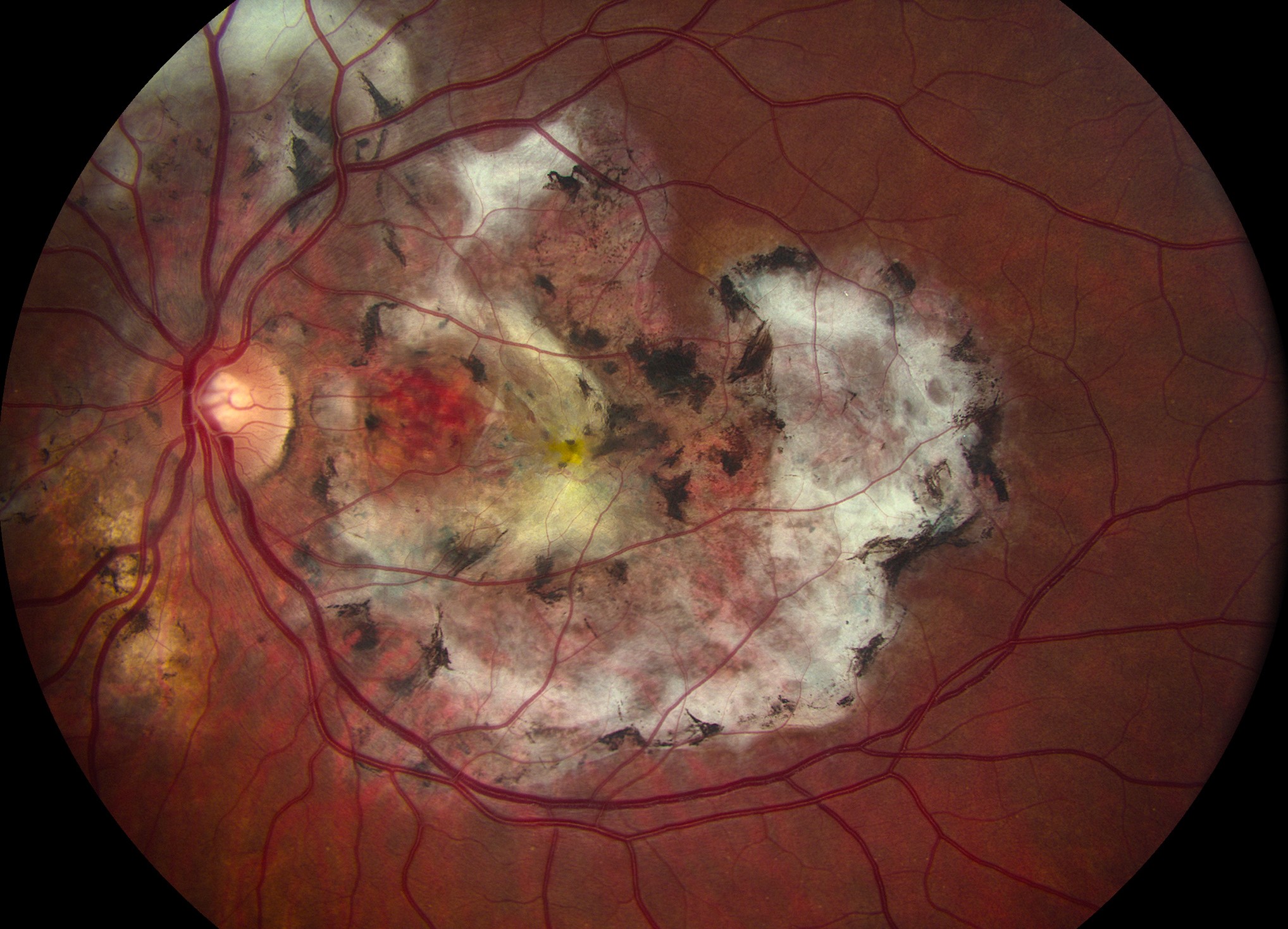
Histopathology of Pneumocystis Choroidopathy
Rao and colleagues examined eyes with the disease at autopsy.
They found an eosinophilic, foamy material in the choroid that contained cysts of the organism.
Also, the infiltrate had little inflammatory cell response, which fits the quiet vitreous seen in life.
The overlying retina was largely spared.
Special stains such as Gomori methenamine silver highlight the cyst walls.
Pneumocystis Choroidopathy in the Era of Antiretroviral Therapy
Shami and colleagues described the multicenter experience in the early epidemic.
Since then, antiretroviral therapy has transformed the course of HIV disease.
As a result, patients with the choroidopathy now are usually newly diagnosed or not adherent to treatment.
Also, consider it in patients on immunosuppression after transplant or chemotherapy.
Therefore, keep it on the list when multifocal yellow lesions appear in any immunocompromised host.
Practical Points for the Clinician
Examine every patient with advanced HIV with dilation, even when vision is normal.
Also take photographs, so that response to treatment can be shown.
Because the lesions are asymptomatic, they are easy to miss without a deliberate search.
Then discuss the finding with the infectious disease team on the same day.
Why the Choroid Is Involved
The choroid has a rich blood supply and sluggish flow, so organisms from the blood settle there.
Also, the retinal pigment epithelium protects the retina from the infection.
As a result, vision stays good while the choroid carries the burden.
This explains the quiet appearance of the vitreous and the retina.
Key Points for the Clinician
Multifocal yellow choroidal lesions in advanced HIV point to Pneumocystis until proven otherwise.
Also, the finding signals systemic disease that needs urgent treatment.
Because the eye is a window, early recognition can save the patient’s life.
Therefore, always arrange a full medical evaluation.
Counselling the Patient
Explain that the eye lesions reflect an infection elsewhere in the body.
Also tell the patient that treatment usually clears the lesions and protects vision.
Because adherence to antiretroviral therapy prevents recurrence, discuss support options.
For the latest studies, see the PubMed results for Pneumocystis choroidopathy.


Document what you see
Two smartphone imaging tools built for everyday clinic use — one for the slit lamp, one for the fundus.
From Choroida — the team behind this siteReferences
- Freeman WR, Gross JG, Labelle J, et al. Pneumocystis carinii choroidopathy: a new clinical entity. Arch Ophthalmol. 1989;107:863-867.
- Rao NA, Zimmerman PL, Boyer D, et al. A clinical, histopathologic, and electron microscopic study of Pneumocystis carinii choroiditis. Am J Ophthalmol. 1989;107:218-228.
- Dugel PU, Rao NA, Forster DJ, et al. Pneumocystis carinii choroiditis after long-term aerosolized pentamidine therapy. Am J Ophthalmol. 1990;110:113-117.
- Shami MJ, Freeman W, Friedberg D, et al. A multicenter study of Pneumocystis choroidopathy. Am J Ophthalmol. 1991;112:15-22.

Writes and reviews the clinical articles on Educate. Every post is checked against peer-reviewed sources before it goes live.