Dominant cystoid macular dystrophy is a rare autosomal dominant disorder in which cystic spaces develop in both maculas without an acquired cause.
The disease starts in young adulthood and causes slowly progressive loss of central vision.
It must be separated from acquired cystoid edema and from other inherited maculopathies.
What Dominant Cystoid Macular Dystrophy Is
Deutman and colleagues described a family with inherited cystoid macular edema in 1976.
Fishman and colleagues later reported additional families with a similar dominant pattern.
The condition is distinct from retinoschisis and from acquired edema, though it can look alike on OCT (see cystoid macular edema).
Clinical Features of Dominant Cystoid Macular Dystrophy
- Bilateral cystic changes in the central macula with a petaloid arrangement
- Onset in the second to fourth decade, often with mild distortion
- Slow decline in acuity, with atrophy in older patients
- No history of uveitis, diabetes, vascular occlusion, or surgery
The family history shows a dominant pattern with affected members in every generation.
Fundus Explorer Pro
Photograph the retinal findings described here with the phone already in your pocket — 22 D optics and built-in illumination in one handheld unit.
From Choroida — the team behind this siteInvestigations in Dominant Cystoid Macular Dystrophy
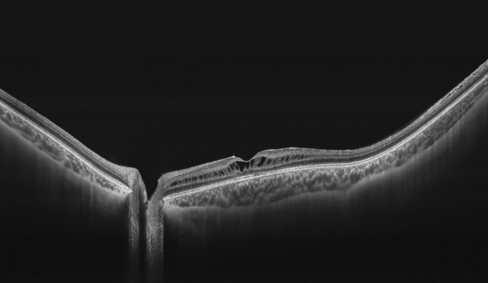
OCT shows cystic spaces in the inner and outer nuclear layers.
Fluorescein angiography may show a petaloid pattern with limited leakage in some patients, so reports vary.
Electrophysiology often shows normal full-field ERG with an abnormal EOG or macular function in some cases.
Genetic testing may find a pathogenic variant, though the genetics remain incompletely defined (see occult macular dystrophy).
Differential Diagnosis of Dominant Cystoid Macular Dystrophy
- X-linked retinoschisis in young males
- Nicotinic acid maculopathy and other drug effects
- Retinitis pigmentosa with cystoid edema
- Uveitic and vascular macular edema
The history and the family pattern usually separate these (see X-linked retinoschisis).
Management of Dominant Cystoid Macular Dystrophy
No proven therapy exists.
Carbonic anhydrase inhibitors have helped some patients with inherited cystoid changes, so consider a trial.
Provide genetic counseling and low vision support as needed.
Prognosis of Dominant Cystoid Macular Dystrophy
The course is slow, and many patients keep reading vision into middle age.
Later atrophy may reduce acuity to 20/200 in some families.
An Example Case
A 24-year-old woman has gradual blurring in both eyes, with acuity of 20/40.
OCT shows symmetric cystic spaces in both maculas, and she has no history of uveitis or diabetes.
Her father and a paternal aunt have similar vision loss.
Genetic testing and family study support a dominant cystoid macular dystrophy, and the team starts a trial of topical dorzolamide.
Questions Patients Ask
Patients ask whether the cysts can be drained, and surgery is not helpful in this condition.
They also ask whether their children will have it, and each child has a 50 percent chance of inheriting the gene.
Because the course is slow, explain that most patients keep useful vision for years.
Pitfalls to Avoid
- Treating the cysts as diabetic or uveitic edema without checking the history
- Skipping the family history in a young patient with bilateral cysts
- Forgetting that retinoschisis can look similar on OCT
- Promising a drug response before a trial is done
Looking Ahead
Genetic studies may identify the responsible genes and clarify the biology.
Also, trials of carbonic anhydrase inhibitors may define their role.
Therefore, patients may soon have better guidance on treatment.
Practical Points
Record the family tree and examine relatives when possible.
Then document OCT thickness at baseline and at each visit.
Also test for other causes of cystoid change.
Imaging Follow-Up
OCT shows the cystic spaces and the retinal thickness over time.
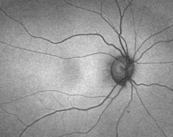
Autofluorescence shows RPE changes in later stages.
Because progression is slow, an annual visit is enough in most patients.
Counselling on Heredity
Explain autosomal dominant inheritance with plain examples.
Then offer to test or examine at-risk relatives.
Also discuss pregnancy planning when asked.
Genetic Counselling
Explain the dominant pattern, with a 50 percent chance for each child.
Then offer testing to relatives when a gene is known.
Because the disease may start in the teens, check at-risk children in their early years.
Also discuss that expression may vary within a family.
Medical Therapy Trials
Topical or oral carbonic anhydrase inhibitors lower cystic spaces in some patients with inherited edema.
However, response differs between families, and relapse may follow when treatment stops.
Therefore, monitor OCT after starting a drug and keep the lowest effective dose.
Also check kidney function and electrolytes during oral therapy.
Summary
Think of dominant cystoid macular dystrophy in a young patient with bilateral cysts and a family history.
Then exclude acquired causes and test the family.
Because the course is slow, plan long-term follow-up.
For the latest studies, see the PubMed results for dominant cystoid macular dystrophy.


Document what you see
Two smartphone imaging tools built for everyday clinic use — one for the slit lamp, one for the fundus.
From Choroida — the team behind this siteReferences
- Deutman AF, Pinckers AJ, Aan de Kerk AL. Dominantly inherited cystoid macular edema. Am J Ophthalmol. 1976;82:540-548.
- Fishman GA, Goldberg MF, Trautmann JC. Dominantly inherited cystoid macular edema. Ann Ophthalmol. 1979;11:21-27.
- Salmon JF. Kanski’s Clinical Ophthalmology: A Systematic Approach. 9th ed. Elsevier; 2020.
- American Academy of Ophthalmology. Basic and Clinical Science Course, Section 12: Retina and Vitreous. AAO; current edition.

Writes and reviews the clinical articles on Educate. Every post is checked against peer-reviewed sources before it goes live.