Macular translocation is a surgical method that moves the fovea away from a subfoveal choroidal neovascular lesion onto healthier retinal pigment epithelium.
It appeared in the 1990s, when treatment of subfoveal neovascular AMD was limited.
Anti-VEGF drugs have since replaced it, but it still shows how ingenious retinal surgery can be.
Why Macular Translocation Was Developed
Subfoveal choroidal neovascularization destroyed central vision, and laser could not treat it without harming the fovea.
Surgeons reasoned that if the fovea moved onto healthy tissue, the neovascular membrane could be treated with laser or removed.
The method needed a healthy pigment epithelium beneath the new fovea (see macular neovascularization types).
Techniques of Macular Translocation
Two main techniques exist.
- 360-degree retinotomy with rotation, described by Machemer and Steinhorst
- Limited macular translocation, in which the surgeon detaches the retina and shifts it by scleral shortening
In the 360-degree method, the surgeon rotates the retina around the optic nerve after a full retinotomy, then applies laser and tamponade.
Meanwhile, the limited version avoids the retinotomy and reduces trauma, and de Juan reported early results.
Fundus Explorer Pro
Photograph the retinal findings described here with the phone already in your pocket — 22 D optics and built-in illumination in one handheld unit.
From Choroida — the team behind this siteTechnique in Brief
The surgeon performs a vitrectomy and creates a subretinal bleb with fluid.
In the 360-degree approach, the surgeon cuts the retina circumferentially and rotates it, then reattaches it with perfluorocarbon and silicone oil.
In the limited approach, the surgeon shortens the sclera or uses a sutured fold to slide the retina.
Eckardt and colleagues added counter-rotation of the globe to correct torsional diplopia (see pars plana vitrectomy).
Results of Macular Translocation
Reports described improved vision in some eyes, with a relatively high rate of reading acuity in selected patients.
However, results varied, and the recurrence of neovascularization often limited the gain.
Comparative trials against photodynamic therapy suggested modest benefits, with more complications.
Complications of Macular Translocation
- Retinal detachment and proliferative vitreoretinopathy
- Torsional diplopia, since the retina rotates
- Macular pucker and hole
- Reoperation in many eyes
These risks explain why most surgeons abandoned the procedure.
Why Anti-VEGF Replaced It
Anti-VEGF injections improved vision without surgery in many eyes, and they carried a lower risk.
Landmark trials such as MARINA and ANCHOR changed the standard of care after 2006 (see anti-VEGF therapy).
Therefore, macular translocation now has little role, apart from rare refractory cases.
An Example Case
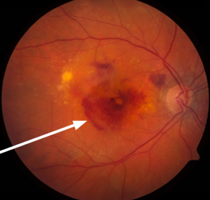
A 62-year-old woman had a subfoveal CNV with vision of 20/200 before anti-VEGF drugs existed.
She chose a limited macular translocation, and the surgeon moved the fovea onto healthy RPE.
Vision improved to 20/80, but she needed surgery for diplopia a year later.
Key Points
- Translocation moves the fovea off the lesion
- It carries high complication rates
- Anti-VEGF therapy has replaced it
Questions Patients Ask
Patients ask whether the surgery is still an option, and the answer is that it is rarely offered today.
They also ask about alternatives, and anti-VEGF injections work better in most eyes.
Because the surgery carries high risk, only expert centers perform it, if at all.
Pitfalls to Avoid
- Operating without a healthy RPE under the new fovea
- Ignoring torsional diplopia before surgery
- Overlooking PVR risk in eyes with large lesions
- Underestimating the need for repeat surgery
Looking Ahead
Interest in retinal rotation continues in rare refractory cases and in research on RPE transplantation.
Also, RPE patch transplantation may replace the need for translocation.
However, these methods remain experimental, so anti-VEGF drugs stay the first choice.
Historical Value
The technique taught surgeons about retinal reattachment and RPE health.
Therefore, it still informs modern work on subretinal surgery and cell therapy.
Patient Selection in the Translocation Era
Surgeons looked for eyes with a subfoveal lesion, a healthy surrounding RPE, and good potential acuity.
They also excluded eyes with extensive atrophy, severe myopia, or scarring.
Because the procedure was long and risky, patients needed strong motivation and a realistic view of the results.
Then they needed careful counseling about diplopia, which often required later surgery.
Photodynamic therapy was the main alternative at that time, with modest benefit.
Even in expert hands, the surgery needed long operating times and careful postoperative care.
Surgeons also had to manage the cyclotorsion that followed rotation.
Therefore, many patients needed a second procedure to realign the images.
For the latest studies, see the PubMed results for macular translocation.


Document what you see
Two smartphone imaging tools built for everyday clinic use — one for the slit lamp, one for the fundus.
From Choroida — the team behind this siteReferences
- Machemer R, Steinhorst UH. Retinal separation, retinotomy, and macular relocation: II. A surgical approach for age-related macular degeneration? Graefes Arch Clin Exp Ophthalmol. 1993;231:635-641.
- De Juan E Jr, Loewenstein A, Bressler NM, Alexander J. Translocation of the retina for management of subfoveal choroidal neovascularization II: a preliminary report in humans. Am J Ophthalmol. 1998;125:635-646.
- Eckardt C, Eckardt U, Conrad HG. Macular rotation with and without counter-rotation of the globe in patients with age-related macular degeneration. Graefes Arch Clin Exp Ophthalmol. 1999;237:313-325.
- American Academy of Ophthalmology. Basic and Clinical Science Course, Section 12: Retina and Vitreous. AAO; current edition.

Writes and reviews the clinical articles on Educate. Every post is checked against peer-reviewed sources before it goes live.