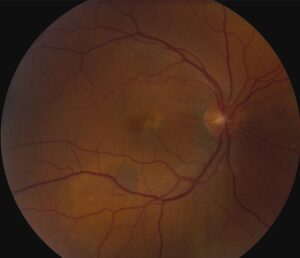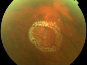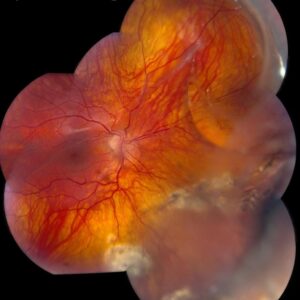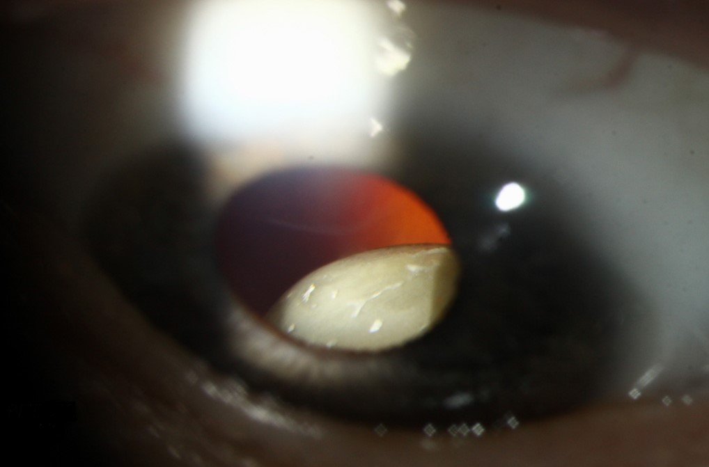
Ectopia lentis means that the crystalline lens sits away from its normal central position because the zonules are weak, stretched, or broken.
A subluxated lens remains partly in the pupillary area, while a luxated lens has moved completely out of it, into the vitreous or the anterior chamber.
The cause may also be inherited, systemic, or acquired, and finding it often leads to a diagnosis that matters for the whole body.
Recognizing Ectopia Lentis
Children usually present with blurred vision, high or variable astigmatism, monocular diplopia through the edge of the lens, or poor response to glasses.
At the slit lamp, look for iridodonesis, phacodonesis.
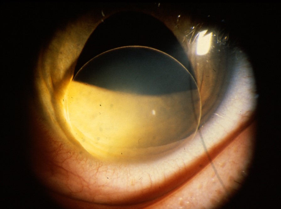
The lens equator crossing the dilated pupil, with the displacement direction recorded for each eye (see slit lamp examination).
Bilateral and symmetric displacement points to a systemic or genetic cause, and unilateral displacement suggests trauma.
Ectopia Lentis: Causes and the Direction of Displacement
- Marfan syndrome: FBN1 mutations, with superotemporal displacement in most patients (see ectopia lentis in Marfan syndrome)
- Homocystinuria: inferonasal displacement, thromboembolic risk, and developmental delay (see homocystinuria)
- Weill-Marchesani syndrome: small, round lenses with inferior displacement and angle closure (see Weill-Marchesani syndrome)
- Isolated and ADAMTSL4 forms: including ectopia lentis et pupillae (see ectopia lentis et pupillae)
- Acquired: blunt trauma, pseudoexfoliation, uveal tumors, and buphthalmos
Sulfite oxidase deficiency, hyperlysinemia, and Ehlers-Danlos syndrome are less common causes.
All-fit Slit-Lamp Adapter
Record and share exactly what you see at the slit lamp. One adapter fits any slit lamp or surgical microscope — and any smartphone.
From Choroida — the team behind this siteSystemic Evaluation
Marfan syndrome carries a risk of aortic root dilatation and dissection, so an echocardiogram is part of the workup.
The revised Ghent criteria give weight to lens displacement (see the references).
Homocystinuria needs plasma homocysteine and amino acid testing, because early treatment with pyridoxine, diet, and betaine reduces thrombotic events.
General anesthesia in these patients should be planned with the anesthetist, since homocystinuria raises the risk of thrombosis.
Complications of Ectopia Lentis
The lens can cause refractive errors, amblyopia, cataract, pupillary block with angle closure, and retinal detachment.
Luxation into the anterior chamber damages the endothelium.
A lens trapped in the pupil can also raise the pressure within hours.
Retinal detachment is more common in Marfan syndrome, so patients need a peripheral retinal examination at regular intervals.
Nonsurgical Management for Ectopia Lentis
Many eyes do well with correction.
Try first a phakic correction through the lens, or an aphakic correction through the edge of the pupil, whichever gives the best acuity.
Treat amblyopia early with patching, and review the refraction often because lens position and astigmatism change.
Surgical Management
Doctors advise surgery when correction cannot restore vision, the lens touches the cornea, pupillary block or glaucoma develops, or a cataract blocks vision.
Lensectomy through the pars plana or a limbal approach with capsular support and a capsular tension ring are also used. The choice depends on how much zonular support remains (see capsular tension ring).
Intraocular lens options include scleral or iris fixation, an anterior chamber lens, or leaving the eye aphakic with a contact lens (see scleral fixated intraocular lens).
Follow-Up
Children need lifelong monitoring of the refraction, the pressure, and the retinal periphery.
In addition, examine siblings and parents, and genetic counselling helps families to plan.
An Example Case
A 7-year-old boy fails a school vision screening, and his glasses never give better than 20/60.
He is tall with long fingers, and the lens edge is visible in the superotemporal pupil of both eyes, with a mild myopic astigmatism that changes at each visit.
An echocardiogram shows a mildly dilated aortic root, and genetic testing confirms an FBN1 mutation, so he starts cardiac follow-up.
The optometrist adjusts his glasses through the lens edge.
Questions Families Ask
Parents ask whether the lens will fall back.
It may move further over time, and the plan is to monitor, correct the vision, and operate only when needed.
They also ask about sports. Advise them to avoid contact sports or trampolines in Marfan syndrome and in eyes with weak zonules, because of the risk to the lens, the retina, and the aorta.
Tell parents to report sudden pain, redness, or loss of vision at once, since an acute pupillary block needs urgent care.
Looking Ahead
Better genotype and phenotype correlations are helping to predict which children will need lens surgery early.
Newer techniques with capsular support devices and sutureless scleral fixation are expanding options for eyes with no zonular support, and registries are collecting long-term results.
When to Refer
Refer any child with unexplained lens displacement to a pediatric ophthalmologist and a geneticist. Refer any adult with sudden displacement to a retina specialist for an examination of the periphery.


Document what you see
Two smartphone imaging tools built for everyday clinic use — one for the slit lamp, one for the fundus.
From Choroida — the team behind this siteReferences
- Nelson LB, Maumenee IH. Ectopia lentis. Surv Ophthalmol. 1982;27:143-160.
- Loeys BL, Dietz HC, Braverman AC, et al. The revised Ghent nosology for the Marfan syndrome. J Med Genet. 2010;47:476-485.
- Chandra A, Aragon-Martin JA, Hughes K, et al. A genotype-phenotype comparison of ADAMTSL4 and FBN1 in isolated ectopia lentis. Ophthalmology. 2012;119:2050-2056.
- American Academy of Ophthalmology. Basic and Clinical Science Course, Section 11: Lens and Cataract. AAO; current edition.

Writes and reviews the clinical articles on Educate. Every post is checked against peer-reviewed sources before it goes live.