White-eyed blowout is a trapdoor fracture of the orbital floor, usually in a child, with entrapment of the inferior rectus or its surrounding tissue.
The eye often looks almost normal at first glance, but the trapped muscle can become ischemic within hours. As a result, this injury needs early recognition and prompt surgical release.
What Is a White-Eyed Blowout?
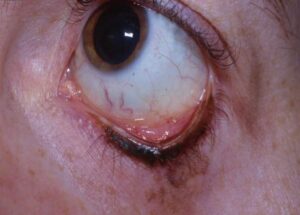
Jordan and colleagues popularized the term in 1998 for floor fractures with few external soft tissue signs. The conjunctiva stays white and lid ecchymosis may be minimal, yet the patient shows marked restriction of vertical gaze. This mismatch between a quiet eye and a severe motility deficit defines the entity.
Why Children Get Trapdoor Fractures
Pediatric bone is elastic and less mineralized than adult bone, so a blow to the orbit cracks the floor, which then snaps back into place. Herniated muscle or fat becomes trapped as the fragment closes like a hinge. In adults, the brittle floor usually shatters and leaves an open defect (see adult orbital blowout fracture).
All-fit Slit-Lamp Adapter
Record and share exactly what you see at the slit lamp. One adapter fits any slit lamp or surgical microscope — and any smartphone.
From Choroida — the team behind this siteTypical History in White-Eyed Blowout
Most cases follow a ball, fist, elbow or fall injury to the orbit, and the child often complains of pain on attempted upgaze. Nausea and vomiting appear early and may dominate the picture, so emergency staff can mistake the pattern for a head injury.
Examination Signs in White-Eyed Blowout
The globe usually looks white, with little subconjunctival hemorrhage. Then motility testing reveals the problem.
- Marked limitation of upgaze, downgaze or both
- Pain on attempted vertical movement
- Little periorbital edema or ecchymosis
- Bradycardia, nausea or faintness during eye movement
The Oculocardiac Reflex
Traction on the trapped muscle stimulates the trigeminal nerve, and the signal then reaches the vagal nucleus and slows the heart. As a result, the child may develop bradycardia, nausea, vomiting or heart block. Because these signs persist until release, many surgeons treat them as a surgical indication in their own right.
Why Imaging Can Mislead
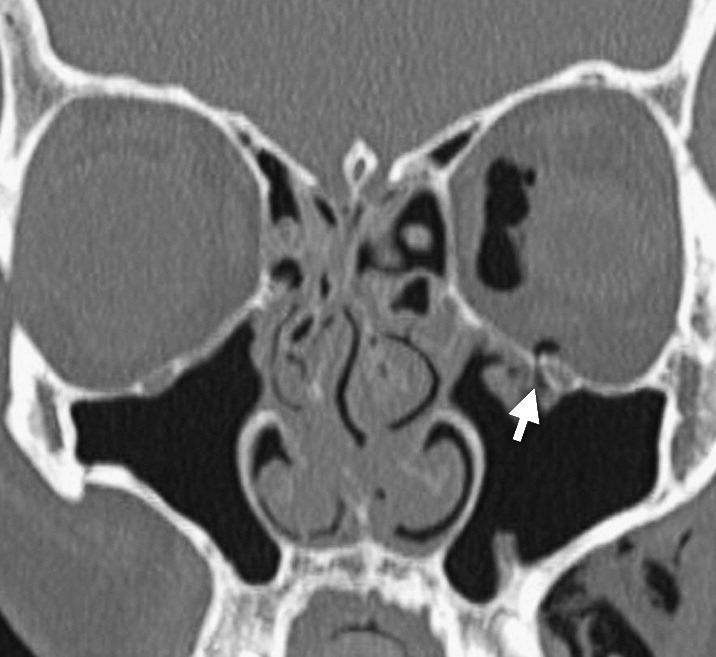
Thin-cut orbital CT with coronal views is the study of choice, but the fracture line may be tiny or barely displaced. A report may therefore describe a normal scan or only a small crack. For this reason, a convincing clinical picture should outweigh a reassuring report.
Missing Muscle Sign in White-Eyed Blowout
Coronal images normally show the inferior rectus resting just above the floor, but in a trapdoor fracture the muscle may appear absent from its usual position. This “missing rectus” appearance suggests the muscle sits within the fracture, so look for a small tuft of soft tissue hanging into the maxillary sinus.
Forced Duction Testing
Under topical anesthesia, the examiner grasps the conjunctiva near the inferior limbus and rotates the globe upward. Resistance confirms a mechanical restriction and separates it from paresis. In young children, cooperation is poor, so the test often takes place under general anesthesia, and clinical restriction with a compatible CT usually justifies proceeding.
Why Surgery in White-Eyed Blowout Is Urgent
The fragment squeezes the trapped muscle and cuts its blood supply, and ischemia then leads to fibrosis and permanent restriction. Several series linked delayed release with persistent motility deficits and diplopia. Therefore, many surgeons aim to operate within the first day or two, and sooner with oculocardiac signs.
Surgical Approach
A transconjunctival or subciliary incision exposes the floor, and the surgeon then frees the incarcerated muscle and fat from the fracture edges. Sometimes the surgeon gently lifts or trims the fragment to release tissue, and a thin implant may cover the defect if it is unstable. Finally, a forced duction test confirms free movement before closure.
Counseling the Family
Parents often struggle to accept urgent surgery for an eye that looks fine, so explain that the danger lies in the muscle, which cannot be seen from outside. Also warn about swelling, temporary double vision and cheek numbness, and finally mention that a second procedure is sometimes needed.
White-Eyed Blowout Versus Adult Blowout Fracture
Adult floor fractures usually allow observation for one to two weeks, during which swelling settles and surgeons then judge enophthalmos and diplopia. In contrast, the white-eyed blowout follows a much shorter clock, and waiting for edema to subside wastes the window for muscle recovery.
Follow-Up After White-Eyed Blowout Repair
Review the child within the first week and then over the following months, check ductions, alignment in all gazes and globe position, and also assess amblyopia risk in younger children with a persistent deviation.
Pitfalls to Avoid
- Do not attribute vomiting to concussion before testing eye movements
- Never trust a “normal” CT when upgaze is severely restricted
- Avoid waiting for swelling to settle in a child with entrapment
- Watch the heart rate during motility testing


Document what you see
Two smartphone imaging tools built for everyday clinic use — one for the slit lamp, one for the fundus.
From Choroida — the team behind this siteReferences
- Jordan DR, Allen LH, White J, Harvey J, Pashby R, Esmaeli B. Intervention within days for some orbital floor fractures: the white-eyed blowout. Ophthalmic Plast Reconstr Surg. 1998;14:379-390.
- Egbert JE, May K, Kersten RC, Kulwin DR. Pediatric orbital floor fracture: direct extraocular muscle involvement. Ophthalmology. 2000;107:1875-1879.
- Grant JH 3rd, Patrinely JR, Weiss AH, Kierney PC, Gruss JS. Trapdoor fracture of the orbit in a pediatric population. Plast Reconstr Surg. 2002;109:482-489.
- Bansagi ZC, Meyer DR. Internal orbital fractures in the pediatric age group: characterization and management. Ophthalmology. 2000;107:829-836.
- Burnstine MA. Clinical recommendations for repair of isolated orbital floor fractures: an evidence-based analysis. Ophthalmology. 2002;109:1207-1210.
Test yourself
A few questions straight from this article.
-
Which mismatch defines a white-eyed blowout fracture of the orbital floor?
The conjunctiva stays white and lid ecchymosis may be minimal, yet vertical gaze is markedly restricted; this mismatch defines the entity. -
Why do children develop trapdoor orbital floor fractures rather than open defects?
Pediatric bone is elastic and less mineralized, so the floor cracks and snaps back like a hinge, trapping muscle or fat; adult floors usually shatter. -
In a child with a white-eyed blowout, early nausea and vomiting are often mistaken for which condition?
Nausea and vomiting appear early and may dominate, so staff can mistake the pattern for head injury; test eye movements before blaming concussion. -
In white-eyed blowout, traction on the trapped muscle slows the heart through which pathway?
The oculocardiac reflex runs from the trigeminal nerve to the vagal nucleus, causing bradycardia, nausea, vomiting or heart block until the muscle is released. -
How should a reassuring orbital CT report be weighed in a child with severe upgaze restriction after orbital trauma?
On thin-cut coronal CT the fracture line may be tiny or barely displaced, so a convincing clinical picture outweighs a reassuring report. -
What does the 'missing rectus' appearance on coronal CT suggest in a trapdoor fracture?
The inferior rectus normally rests just above the floor; when it appears absent, it likely sits within the fracture, sometimes as a soft-tissue tuft in the sinus. -
In suspected white-eyed blowout, what does resistance on forced duction testing confirm?
Grasping the conjunctiva near the inferior limbus and rotating the globe upward meets resistance in mechanical restriction, separating it from paresis. -
Within what time frame do many surgeons aim to release a white-eyed blowout fracture?
Many surgeons aim to operate within the first day or two, and sooner with oculocardiac signs, unlike adult floor fractures that allow observation. -
Why does delayed release of a white-eyed blowout risk permanent motility restriction?
The fragment squeezes the trapped muscle and cuts its blood supply; ischemia then causes fibrosis, and delayed release has been linked to persistent diplopia. -
After white-eyed blowout repair, what should be assessed in a younger child with a persistent deviation?
Follow-up checks ductions, alignment and globe position, and assesses amblyopia risk in younger children with a persistent deviation.

Writes and reviews the clinical articles on Educate. Every post is checked against peer-reviewed sources before it goes live.