Eye discharge is any material that collects at the lid margin, lashes or inner canthus.
Its texture, color and timing often point to the cause before the slit lamp exam. However, discharge type supports a diagnosis and never replaces a full examination.
What Counts as Eye Discharge
Normal tears carry a little mucus, which dries into crust overnight, so many healthy people wake with “sleep” in the inner corners. Pathologic eye discharge differs in amount, color, persistence or speed of reaccumulation, so also ask whether discharge affects one or both eyes and how quickly it returns after wiping.
The Main Types of Eye Discharge
Clinicians usually sort discharge into five patterns.
- Watery (serous): clear fluid, often with follicles
- Mucoid: thin, clear or white mucus strands
- Ropy: thick, elastic mucus that stretches when lifted
- Mucopurulent: yellow or green, sticky, gluing the lashes
- Hyperpurulent: copious pus that returns within minutes
All-fit Slit-Lamp Adapter
Record and share exactly what you see at the slit lamp. One adapter fits any slit lamp or surgical microscope — and any smartphone.
From Choroida — the team behind this siteWatery Eye Discharge
Serous discharge is typical of viral conjunctivitis, especially adenovirus. Look also for a tender preauricular node, follicles and later subepithelial infiltrates, because toxic reactions to drops and mild allergy can also produce watery eyes.
With pain or photophobia, however, suspect keratitis, a foreign body or anterior uveitis. Tearing without inflammation points instead to the lacrimal drainage system (see watery eye in adults).
Mucoid Eye Discharge
Thin mucus strands suggest allergic conjunctivitis or dry eye, and itching favors allergy, while burning and grittiness favor tear film disease. Some dry eye patients repeatedly pull mucus from the fornix, and as a result, mechanical trauma increases mucus production and the cycle continues.
Ropy Mucus and Allergy
Thick, stringy, elastic mucus is the hallmark of vernal and atopic keratoconjunctivitis, so evert the upper lid and look for giant papillae. Also check the limbus for Horner-Trantas dots and the cornea for shield ulcers, because shield ulcers threaten vision and active disease needs prompt specialist care.
Mucopurulent Eye Discharge
Yellow discharge with lashes glued together suggests acute bacterial conjunctivitis, and common organisms include Staphylococcus aureus, Streptococcus pneumoniae and Haemophilus influenzae. In one primary care cohort, both eyes glued in the morning favored a bacterial cause, while itching, by contrast, argued against it. However, adenoviral conjunctivitis can also turn mucopurulent, so overlap is common.
Antibiotics or Watchful Waiting
Acute bacterial conjunctivitis often settles without treatment, although a Cochrane review found that topical antibiotics modestly speed early remission. Therefore, delayed prescribing is a reasonable option for mild cases in adults, and you should advise hand hygiene and separate towels, because household spread is common.
Chronic Mucopurulent Discharge
Discharge lasting beyond about four weeks needs a broader differential, so first consider adult chlamydial conjunctivitis, especially with follicles and a palpable node. Also consider molluscum on the lid margin, canaliculitis and dacryocystitis, and finally, chronic unilateral “conjunctivitis” in an older adult can mask sebaceous carcinoma.
Hyperpurulent Eye Discharge
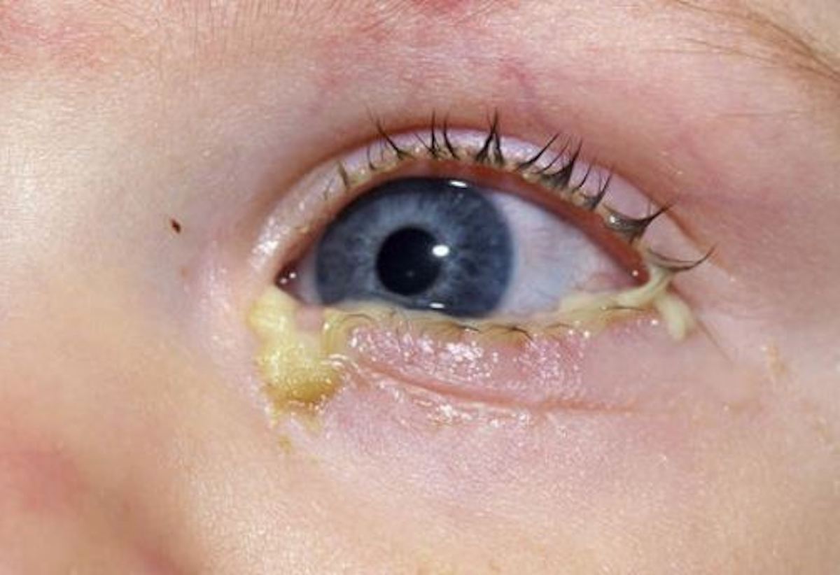
Copious pus that returns minutes after wiping is an emergency, and Neisseria gonorrhoeae is the classic cause, with marked chemosis and lid swelling. This organism can invade intact corneal epithelium, and perforation may follow quickly. Therefore, take swabs for Gram stain and culture, then start systemic ceftriaxone, and also irrigate the fornices and test for chlamydial coinfection.
Eye Discharge in Newborns
Discharge in the first month raises concern for ophthalmia neonatorum, and gonococcal disease tends to appear within days, while chlamydia usually appears later. Both need urgent swabs and systemic treatment, but in contrast, a quiet eye with sticky discharge often reflects nasolacrimal duct obstruction.
Lacrimal Sources of Pus
Press over the lacrimal sac and watch the puncta, where reflux of mucopus indicates nasolacrimal obstruction, often with dacryocystitis. A pouting, inflamed punctum with yellow granules suggests canaliculitis, often from Actinomyces, and because the source lies in the canaliculus, repeated drops rarely cure it.
Lid Margin Crusting
Crusts at the lash base usually reflect blepharitis, which patients often call discharge. Look for collarettes, capped meibomian glands and lid margin telangiectasia, and daily lid hygiene helps, although symptoms tend to recur.
Contact Lenses and Eye Discharge
Any discharge in a contact lens wearer needs a careful corneal exam, because Pseudomonas keratitis can begin with mucopurulent discharge and rapid pain. Therefore, stop lens wear and search for an infiltrate (see keratitis vs conjunctivitis).
Red Flags in Eye Discharge
- Reduced vision or a corneal infiltrate
- Moderate or severe pain and photophobia
- Hyperpurulent discharge
- Any discharge in a neonate
- Contact lens wear with a painful red eye
- Lid swelling with fever, proptosis or painful eye movements (see orbital cellulitis red flags)
- Unilateral chronic disease that fails treatment
Pitfalls to Avoid
- Do not start steroid drops without a slit lamp exam
- Never treat hyperpurulent discharge as routine conjunctivitis
- Avoid repeated antibiotic switches without cultures
- Do not miss canaliculitis behind chronic conjunctivitis


Document what you see
Two smartphone imaging tools built for everyday clinic use — one for the slit lamp, one for the fundus.
From Choroida — the team behind this siteReferences
- Azari AA, Barney NP. Conjunctivitis: a systematic review of diagnosis and treatment. JAMA. 2013;310:1721-1729.
- Varu DM, Rhee MK, Akpek EK, et al. Conjunctivitis Preferred Practice Pattern. Ophthalmology. 2019;126:P94-P169.
- Rietveld RP, ter Riet G, Bindels PJ, Sloos JH, van Weert HC. Predicting bacterial cause in infectious conjunctivitis: cohort study on informativeness of combinations of signs and symptoms. BMJ. 2004;329:206-210.
- Sheikh A, Hurwitz B, van Schayck CP, McLean S, Nurmatov U. Antibiotics versus placebo for acute bacterial conjunctivitis. Cochrane Database Syst Rev. 2012;9:CD001211.
- Bowling B. Kanski’s Clinical Ophthalmology: A Systematic Approach. 8th ed. Edinburgh: Elsevier; 2016.
Test yourself
A few questions straight from this article.
-
Watery (serous) eye discharge is most typical of which condition?
Serous discharge is typical of viral conjunctivitis, especially adenovirus, though drop toxicity and mild allergy can also cause watery eyes. -
Thick, ropy, elastic eye discharge should prompt which examination step?
Ropy mucus is the hallmark of vernal and atopic keratoconjunctivitis, so evert the upper lid for giant papillae and check for shield ulcers. -
With mucopurulent eye discharge, which history favoured a bacterial cause in a primary care cohort?
In one primary care cohort, both eyes glued in the morning favoured a bacterial cause, while itching argued against it. -
What effect do topical antibiotics have in acute bacterial conjunctivitis according to a Cochrane review?
Bacterial conjunctivitis often settles alone, and antibiotics modestly speed early remission, so delayed prescribing is reasonable for mild adult cases. -
Chronic unilateral 'conjunctivitis' with discharge in an older adult can mask which tumour?
Discharge beyond about four weeks needs a broader differential, and chronic unilateral conjunctivitis in an older adult can mask sebaceous carcinoma. -
Why is hyperpurulent eye discharge from Neisseria gonorrhoeae an emergency?
Gonococcus can invade intact corneal epithelium and perforation may follow quickly, so swabs are taken and systemic ceftriaxone started. -
In a newborn, a quiet eye with sticky discharge most often reflects which condition?
Neonatal discharge raises concern for ophthalmia neonatorum, but a quiet eye with sticky discharge often reflects nasolacrimal duct obstruction. -
A pouting, inflamed punctum with yellow granules and chronic discharge suggests which diagnosis?
Canaliculitis, often from Actinomyces, gives a pouting punctum with yellow granules, and because the source is in the canaliculus, drops rarely cure it. -
Which keratitis should be excluded when a contact lens wearer presents with eye discharge?
Pseudomonas keratitis can begin with mucopurulent discharge and rapid pain, so lens wear stops and the cornea is searched for an infiltrate. -
Why does mucoid discharge persist in dry eye patients who repeatedly pull mucus from the fornix?
Repeatedly pulling mucus from the fornix causes mechanical trauma that increases mucus production, so the cycle continues.

Writes and reviews the clinical articles on Educate. Every post is checked against peer-reviewed sources before it goes live.