Vitreous hemorrhage grading scores how densely blood fills the vitreous, based on how much fundus detail remains visible.
No single scale is universal, however most clinicians use a simple stepwise system, and the grade then guides imaging, observation and surgical timing.
Why Vitreous Hemorrhage Grading Helps
A grade turns a vague note such as “some blood” into a comparable record, and it also lets another examiner judge clearance using the same landmarks. Therefore, the grade decides whether ophthalmoscopy alone is safe.
A Practical Vitreous Hemorrhage Grading Scale
The version below is common in practice, although wording varies between centers. Write the definitions into your clinic protocol, so every examiner scores the same way.
- Grade 0: clear vitreous, no visible blood
- Mild (grade 1): disc and major vessels clearly seen, fine detail hazy
- Grade 2, moderate: disc or major vessels seen only partly
- Dense (grade 3): red reflex present, but no fundus detail
- Grade 4, total: no red reflex at all
Fundus Explorer Pro
Photograph the retinal findings described here with the phone already in your pocket — 22 D optics and built-in illumination in one handheld unit.
From Choroida — the team behind this siteExamining the Eye for Vitreous Hemorrhage Grading
First, dilate fully, because a small pupil exaggerates apparent density. Next, use indirect ophthalmoscopy, which penetrates blood better than slit lamp lenses, and also look for pigment cells in the anterior vitreous, since they suggest a retinal break.
Mild Hemorrhage: Grade 1
In grade 1 you can usually examine the posterior pole and much of the periphery. As a result, the cause is often visible, such as neovascularization or a flap tear, and scleral depression then completes the peripheral examination.
Moderate Hemorrhage: Grade 2
Grade 2 hides enough retina to miss a peripheral tear or shallow detachment, and blood often settles inferiorly, so the superior retina may clear first. Therefore, combine indirect ophthalmoscopy with B-scan for every hidden area.
Dense Hemorrhage: Grades 3 and 4
At grades 3 and 4, acuity is often hand motions or worse. However, light projection should remain accurate when the retina is attached, while poor projection or a relative afferent pupillary defect suggests detachment or other posterior disease.
Vitreous Hemorrhage Grading and B-scan Use
B-scan shows mobile vitreous echoes and confirms whether the retina is attached (see B-scan ultrasonography). It may also reveal posterior vitreous detachment, flap tears, tumors or a dislocated lens. In dense nondiabetic hemorrhage, many specialists repeat the scan within one to two weeks, because a negative study can miss small breaks.
Location Matters Alongside Density
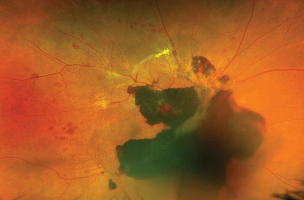
Record where the blood sits, because location changes management. For example, subhyaloid blood forms a boat-shaped level behind the posterior hyaloid, while premacular blood under the internal limiting membrane may need Nd:YAG membranotomy or surgery.
Linking the Grade to the Likely Cause
The grade does not reveal the cause, while history and the fellow eye often do. For example, fellow-eye neovascularization in a diabetic patient points to proliferative retinopathy. In contrast, sudden flashes and floaters in a nondiabetic suggest an acute vitreous detachment with a tear, while other causes include vein occlusion, trauma, neovascular macular degeneration and Terson syndrome.
Observation by Grade
Observation suits grades 1 and 2 when the retina is attached and the cause is known. Also advise head elevation during sleep, because settling blood may clear the visual axis. Anticoagulants usually continue after discussion with the prescribing physician, and laser treatment should then be completed as the view improves.
Anti-VEGF in Diabetic Hemorrhage
In DRCR Protocol N, ranibizumab did not significantly reduce vitrectomy at 16 weeks compared with saline. Later, Protocol AB compared aflibercept with prompt vitrectomy plus panretinal photocoagulation, and it found no significant difference in mean visual acuity over 24 weeks (see Protocol AB). Therefore, injections are a reasonable first option for many diabetic patients.
Vitreous Hemorrhage Grading and Vitrectomy Timing
Dense hemorrhage with a detachment on B-scan needs prompt vitrectomy. The Diabetic Retinopathy Vitrectomy Study found that early surgery benefited type 1 diabetic patients with severe hemorrhage, but that trial predates small-gauge surgery and anti-VEGF therapy. Nonclearing blood, ghost cell glaucoma or a poor fellow eye also favor earlier surgery, yet surgery cannot restore vision lost to macular ischemia.
Tracking Clearance at Follow-up
Record grade, acuity, pressure and B-scan findings at each visit. A falling grade supports observation, while a rising grade suggests rebleeding, and you should also check intraocular pressure, because degenerated red cells can block outflow.
Pitfalls in Vitreous Hemorrhage Grading
- Grading through an undilated pupil overstates density
- Calling a dense bleed “diabetic” without checking for a tear
- Trusting one normal B-scan in a fresh hemorrhage
- Ignoring pressure when blood persists for weeks
- Using local terms that colleagues cannot compare


Document what you see
Two smartphone imaging tools built for everyday clinic use — one for the slit lamp, one for the fundus.
From Choroida — the team behind this siteReferences
- Spraul CW, Grossniklaus HE. Vitreous hemorrhage. Surv Ophthalmol. 1997;42:3-39.
- The Diabetic Retinopathy Vitrectomy Study Research Group. Early vitrectomy for severe vitreous hemorrhage in diabetic retinopathy. Two-year results of a randomized trial. Diabetic Retinopathy Vitrectomy Study report 2. Arch Ophthalmol. 1985;103:1644-1652.
- Diabetic Retinopathy Clinical Research Network. Randomized clinical trial evaluating intravitreal ranibizumab or saline for vitreous hemorrhage from proliferative diabetic retinopathy. JAMA Ophthalmol. 2013;131:283-293.
- Antoszyk AN, Glassman AR, Beaulieu WT, et al. Effect of intravitreous aflibercept vs vitrectomy with panretinal photocoagulation on visual acuity in patients with vitreous hemorrhage from proliferative diabetic retinopathy: a randomized clinical trial. JAMA. 2020;324:2383-2395.
- American Academy of Ophthalmology. Basic and Clinical Science Course, Section 12: Retina and Vitreous. San Francisco: AAO; 2024.
Test yourself
A few questions straight from this article.
-
In a common vitreous hemorrhage grading scale, which finding defines dense (grade 3) hemorrhage?
Grade 3 (dense) hemorrhage keeps a red reflex but shows no fundus detail, whereas grade 4 (total) has no red reflex at all. -
In vitreous hemorrhage grading, what does a moderate (grade 2) hemorrhage allow the examiner to see?
In grade 2 the disc or major vessels are seen only partly, enough retina is hidden to miss a peripheral tear, so indirect ophthalmoscopy is combined with B-scan. -
Why should the pupil be fully dilated before grading a vitreous hemorrhage?
A small pupil exaggerates apparent density, so grading through an undilated pupil overstates the hemorrhage. -
In an eye with vitreous hemorrhage, pigment cells in the anterior vitreous suggest which underlying cause?
During examination for vitreous hemorrhage, pigment cells in the anterior vitreous suggest a retinal break, so the periphery must be examined carefully. -
In dense vitreous hemorrhage, what does poor light projection or a relative afferent pupillary defect suggest?
At grades 3 and 4 acuity is often hand motions, but light projection should stay accurate if the retina is attached; poor projection or an RAPD suggests detachment or other posterior disease. -
Why do many specialists repeat B-scan within one to two weeks in dense nondiabetic vitreous hemorrhage?
B-scan confirms whether the retina is attached, but a negative study can miss small breaks, so the scan is repeated within one to two weeks in dense nondiabetic hemorrhage. -
Premacular blood trapped under the internal limiting membrane may need which treatment?
Location changes management: subhyaloid blood forms a boat-shaped level, while premacular blood under the internal limiting membrane may need Nd:YAG membranotomy or surgery. -
What did DRCR Protocol AB find when comparing aflibercept with prompt vitrectomy plus PRP for diabetic vitreous hemorrhage?
Protocol AB found no significant difference in mean visual acuity over 24 weeks between aflibercept and prompt vitrectomy with panretinal photocoagulation, so injections are a reasonable first option for many diabetic patients. -
Which B-scan finding in dense vitreous hemorrhage calls for prompt vitrectomy?
Dense hemorrhage with a detachment on B-scan needs prompt vitrectomy, while nonclearing blood, ghost cell glaucoma or a poor fellow eye also favour earlier surgery. -
Why should intraocular pressure be checked when vitreous hemorrhage persists for weeks?
At follow-up, pressure is checked because degenerated red cells can block outflow; a falling grade supports observation, while a rising grade suggests rebleeding.

Writes and reviews the clinical articles on Educate. Every post is checked against peer-reviewed sources before it goes live.