Choroidal tumors range from harmless nevi to melanoma, metastasis and lymphoma.
Several of them look alike on a quick fundus view, but color, shape, ultrasound and autofluorescence usually separate them.
Why Choroidal Tumors Look Alike
Clinicians view every choroidal mass through the retinal pigment epithelium. As a result, drusen, lipofuscin and fluid blur the true tissue color, and amelanotic melanoma also loses the brown hue that usually flags malignancy. Therefore, use multimodal imaging, because pattern recognition alone often fails.
Quick Comparison Table of Choroidal Tumors
- Nevus: slate gray, flat, surface drusen; usually under 2 mm on ultrasound
- Melanoma: brown or amelanotic dome or mushroom; acoustic hollowness; bright orange pigment on FAF
- Hemangioma: orange-red dome near the disc; high internal reflectivity
- Osteoma: yellow-orange plaque with scalloped edges; bony plate with shadowing
- Metastasis: creamy yellow, placoid, often multifocal; lipofuscin clumps on FAF
- Lymphoma: salmon-yellow diffuse thickening; low reflectivity, possible extrascleral extension
Fundus Explorer Pro
Photograph the retinal findings described here with the phone already in your pocket — 22 D optics and built-in illumination in one handheld unit.
From Choroida — the team behind this siteColor Clues in Choroidal Tumors
Pigment points toward a melanocytic lesion, although some melanomas are partly or fully amelanotic. Orange-red suggests hemangioma, while yellow-white suggests osteoma or metastasis. A salmon tone, especially with an episcleral patch, raises concern for lymphoma.
Shape and Margins
A flat lesion with feathery edges fits a nevus. In contrast, a dome that breaks Bruch membrane forms the mushroom shape of melanoma. Osteoma stays flat and geographic, while metastases often spread as wide plaques.
Ultrasound in Choroidal Tumors
B-scan measures thickness and shape, while A-scan grades internal reflectivity. Melanoma, for example, typically shows low to medium reflectivity, acoustic hollowness and choroidal excavation.
Hemangioma, by contrast, gives high, regular internal reflectivity, while osteoma produces a dense plate that still shadows the orbit at low gain. Metastasis and lymphoma vary, so ultrasound alone cannot settle them.
Autofluorescence Patterns
Fundus autofluorescence maps the RPE response over the tumor. For example, orange pigment on a pigmented lesion glows brightly and signals growth risk. Drusen on a chronic nevus also glow, but they suggest a stable, long-standing process, and metastases show similar lipofuscin clumps, so FAF supports a diagnosis without proving it.
Choroidal Nevus
Nevi are common and usually stable. However, risk rises with thickness over 2 mm, subretinal fluid or symptoms, so apply the risk factors systematically (see TFSOM risk factors) and photograph and measure every lesion at baseline.
Choroidal Melanoma
Melanoma is the most common primary intraocular malignancy in adults, and it grows, leaks fluid and often carries orange pigment. In COMS, brachytherapy and enucleation gave similar survival for medium tumors. Systemic staging then follows, because liver metastasis drives prognosis.
Circumscribed Hemangioma
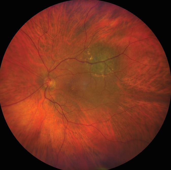
This benign vascular hamartoma usually sits near the optic disc or macula, and patients notice blurred vision from subretinal fluid. ICG angiography shows rapid early filling and late washout, and many surgeons use photodynamic therapy to resolve the fluid.
Choroidal Osteoma
Osteoma mostly affects young women and lies next to the disc, and because it is mature bone, ultrasound and CT both show calcium density. Over time, it may decalcify, and the overlying photoreceptors then atrophy. Next, watch for choroidal neovascularization, which threatens central vision.
Metastasis to the Choroid
Metastasis is the most common intraocular malignancy overall, and breast and lung cancers are the usual primaries. Lesions are often multifocal or bilateral, with heavy fluid for their height, and a notable share of patients have no known cancer at presentation (see choroidal metastases).
Choroidal Lymphoma
Primary choroidal lymphoma is usually a low-grade marginal zone B-cell tumor, and it causes diffuse creamy thickening, sometimes with a salmon episcleral mass. In contrast, vitreoretinal lymphoma causes vitritis and sub-RPE infiltrates (see intraocular lymphoma).
History That Shifts the Diagnosis
First, a known cancer makes metastasis the leading concern, and similarly, Sturge-Weber syndrome points to diffuse hemangioma. Finally, documented growth on serial photographs outweighs any single static feature.
Counseling Patients With Choroidal Tumors
Explain which features look benign and which need watching, then set a clear follow-up interval. Refer suspicious lesions to ocular oncology promptly, and avoid calling a lesion a freckle before imaging is complete.
Pitfalls With Choroidal Tumors
- Do not dismiss an amelanotic lesion because it lacks pigment
- Never skip ultrasound in a lesion thicker than a flat nevus
- Do not confuse osteoma shadowing with melanoma hollowness
- Remember that metastases can also be the first sign of systemic cancer
- Avoid single-visit decisions when growth is the main question


Document what you see
Two smartphone imaging tools built for everyday clinic use — one for the slit lamp, one for the fundus.
From Choroida — the team behind this siteReferences
- Shields CL, Furuta M, Berman EL, et al. Choroidal nevus transformation into melanoma: analysis of 2514 consecutive cases. Arch Ophthalmol. 2009;127:981-987.
- Collaborative Ocular Melanoma Study Group. The COMS randomized trial of iodine 125 brachytherapy for choroidal melanoma, III: initial mortality findings. COMS Report No. 18. Arch Ophthalmol. 2001;119:969-982.
- Shields CL, Shields JA, Gross NE, et al. Survey of 520 eyes with uveal metastases. Ophthalmology. 1997;104:1265-1276.
- Shields CL, Honavar SG, Shields JA, et al. Circumscribed choroidal hemangioma: clinical manifestations and factors predictive of visual outcome in 200 consecutive cases. Ophthalmology. 2001;108:2237-2248.
- Shields CL, Sun H, Demirci H, Shields JA. Factors predictive of tumor growth, tumor decalcification, choroidal neovascularization, and visual outcome in 74 eyes with choroidal osteoma. Arch Ophthalmol. 2005;123:1658-1666.
- Coupland SE, Damato B. Understanding intraocular lymphomas. Clin Exp Ophthalmol. 2008;36:564-578.
Test yourself
A few questions straight from this article.
-
Why do different choroidal tumors often look alike on a quick fundus view?
Every choroidal mass is viewed through the retinal pigment epithelium, where drusen, lipofuscin and fluid blur the true tissue colour, so multimodal imaging is needed. -
What produces the mushroom shape of a choroidal melanoma?
A flat lesion with feathery edges fits a nevus, whereas a dome that breaks Bruch membrane forms the mushroom shape of melanoma. -
Which ultrasound pattern is typical of choroidal melanoma?
Melanoma typically shows low to medium internal reflectivity, acoustic hollowness and choroidal excavation, whereas hemangioma gives high regular reflectivity and osteoma a shadowing bony plate. -
What does indocyanine green angiography show in circumscribed choroidal hemangioma?
This benign vascular hamartoma, usually near the disc or macula, shows rapid early filling and late washout on ICG angiography; many surgeons use photodynamic therapy to resolve its fluid. -
Which complication of choroidal osteoma threatens central vision?
Osteoma is mature bone that may decalcify with overlying photoreceptor atrophy, and choroidal neovascularization is the complication that threatens central vision. -
Above what thickness does the risk of a choroidal nevus being a melanoma rise?
Nevi are usually under 2 mm on ultrasound; risk rises with thickness over 2 mm, subretinal fluid or symptoms, so every lesion should be photographed and measured at baseline. -
Which primary cancers most often metastasize to the choroid?
Metastasis is the most common intraocular malignancy overall, with breast and lung as the usual primaries; lesions are often multifocal or bilateral with heavy fluid for their height. -
What drives prognosis in choroidal melanoma?
Brachytherapy and enucleation gave similar survival for medium tumors, and systemic staging follows because liver metastasis drives prognosis. -
What is primary choroidal lymphoma usually, and how may it present?
Primary choroidal lymphoma is usually low-grade marginal zone B-cell, giving diffuse creamy thickening, sometimes with a salmon episcleral mass, unlike vitreoretinal lymphoma, which causes vitritis. -
Which finding outweighs any single static feature when classifying a choroidal tumor?
Documented growth on serial photographs outweighs any single static feature, so single-visit decisions should be avoided when growth is the main question.

Writes and reviews the clinical articles on Educate. Every post is checked against peer-reviewed sources before it goes live.