Cataract with AMD is a common pairing in older patients, because both conditions rise steeply with age.
The surgeon must decide whether the lens or the macula limits vision, and patients also ask whether surgery will worsen their macular degeneration.
Why Cataract With AMD Is a Common Dilemma
Both diseases blur central vision and reduce contrast sensitivity. As a result, patients often cannot tell which problem causes the complaint, and a dense lens also hides macular detail during examination.
Does Surgery Speed AMD Progression?
Pooled data from the Beaver Dam and Blue Mountains studies linked cataract surgery with more late AMD. However, these cohorts had limited macular imaging before surgery.
Later, AREDS report 25 found no clear association between cataract surgery and progression to advanced AMD. Therefore, most clinicians now regard any added risk as small.
Fundus Explorer Pro
Photograph the retinal findings described here with the phone already in your pocket — 22 D optics and built-in illumination in one handheld unit.
From Choroida — the team behind this siteWeighing the Evidence in Cataract With AMD
A Cochrane review found too few randomized trials to settle the question, so observational data carry most of the weight. Also, a dense lens can hide early neovascularization, so some “progression” may be preexisting disease.
Expected Visual Gain
AREDS report 27 found that acuity improved after surgery across AMD severity levels, and eyes with advanced AMD also gained, although final acuity stayed lower.
Patients often notice brighter images and better color. However, the gain depends on how much loss came from the lens.
Predicting Benefit in Cataract With AMD
First, compare lens density with the degree of visual loss. A dense nuclear cataract with mild drusen predicts a good result, whereas foveal geographic atrophy with a mild cataract predicts little gain. Potential acuity tests help, while dense opacities make them unreliable.
Preoperative Imaging in Cataract With AMD
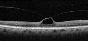
OCT is the most useful test before surgery, and it shows drusen, fluid, atrophy and fibrosis through moderate lens opacity.
Then, autofluorescence defines the extent of geographic atrophy, and a baseline scan also makes postoperative comparison easier.
Timing With Active Neovascular Disease
Control active exudation before elective surgery. Therefore, most retina specialists prefer a dry macula on OCT for a period of stability.
Many surgeons then operate shortly after an injection, within the usual interval. Close coordination with the retina service prevents missed doses (see anti-VEGF therapy).
Anti-VEGF Schedule in Cataract With AMD
Do not pause injections because of cataract surgery. Some surgeons add an injection at the end of surgery, although evidence for this is limited.
After surgery, repeat OCT at the next retina visit, then return the patient to the regular injection plan.
Postoperative Macular Edema
Pseudophakic cystoid macular edema may mimic neovascular reactivation. Because treatment differs, check angiography or OCT angiography when the pattern is unclear. Topical NSAIDs are widely used, although their benefit in AMD is unproven.
Dry AMD and Geographic Atrophy
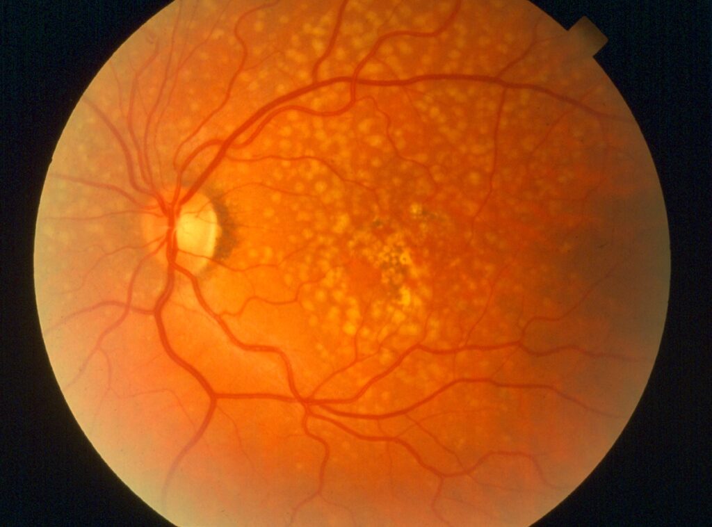
Intermediate dry AMD rarely poses a barrier to surgery. However, foveal atrophy limits the potential gain, but even so, a brighter image can improve eccentric viewing and magnifier use. Atrophy after anti-VEGF therapy needs the same caution (see macular atrophy after anti-VEGF).
IOL Choice in Cataract With AMD
Monofocal lenses remain the standard choice, and multifocal lenses reduce contrast, so avoid them in most eyes with AMD.
Toric lenses are reasonable for significant astigmatism. Blue light filtering lenses have a theoretical rationale. However, no convincing clinical evidence shows that they slow AMD.
When to Operate in Cataract With AMD
Operate when the cataract clearly adds to visual disability. Waiting does not protect the macula, and a clear lens also allows better retinal monitoring and treatment. General timing principles still apply (see cataract surgery timing).
When to Wait
Delay surgery while neovascular disease is active, and also reconsider when vision loss is mainly macular and the lens is mild.
In that case, surgery carries risk with little chance of gain, so low vision referral may help more.
Counseling in Cataract With AMD
First, explain that the macula sets the ceiling for final vision, then describe the likely gain in brightness and contrast.
Patients also need reassurance that evidence does not show clear macular harm. Finally, stress that injections and home Amsler monitoring continue after surgery.
Pitfalls to Avoid
- Operating while exudation is active
- Promising sharp reading vision with foveal atrophy
- Implanting multifocal lenses in eyes with AMD
- Calling all postoperative fluid cystoid macular edema
- Skipping a baseline OCT


Document what you see
Two smartphone imaging tools built for everyday clinic use — one for the slit lamp, one for the fundus.
From Choroida — the team behind this siteReferences
- Chew EY, Sperduto RD, Milton RC, et al. Risk of advanced age-related macular degeneration after cataract surgery in the Age-Related Eye Disease Study: AREDS report 25. Ophthalmology. 2009;116:297-303.
- Forooghian F, Agrón E, Clemons TE, Ferris FL 3rd, Chew EY. Visual acuity outcomes after cataract surgery in patients with age-related macular degeneration: Age-Related Eye Disease Study report no. 27. Ophthalmology. 2009;116:2093-2100.
- Wang JJ, Klein R, Smith W, et al. Cataract surgery and the 5-year incidence of late-stage age-related macular degeneration: pooled findings from the Beaver Dam and Blue Mountains eye studies. Ophthalmology. 2003;110:1960-1967.
- Casparis H, Lindsley K, Kuo IC, Sikder S, Bressler NM. Surgery for cataracts in people with age-related macular degeneration. Cochrane Database Syst Rev. 2017;2:CD006757.
- Mitchell P, Liew G, Gopinath B, Wong TY. Age-related macular degeneration. Lancet. 2018;392:1147-1159.
Test yourself
A few questions straight from this article.
-
What did AREDS report 25 find about cataract surgery and progression to advanced age-related macular degeneration?
Earlier Beaver Dam and Blue Mountains pooled data suggested more late AMD, but those cohorts had limited preoperative macular imaging; AREDS report 25 found no clear association. -
Why may some apparent AMD progression after cataract surgery actually be preexisting disease?
A dense lens can conceal early neovascularization, so disease first seen after surgery may have been present beforehand. -
According to AREDS report 27, how did visual acuity change after cataract surgery in eyes with AMD?
Acuity improved across severity levels, and even eyes with advanced AMD gained, although their final acuity stayed lower. -
In a patient with cataract and AMD, which combination predicts the best visual result from cataract surgery?
Benefit depends on how much loss comes from the lens; a dense nuclear cataract with mild drusen predicts a good result, foveal atrophy with mild cataract little gain. -
Which preoperative test is most useful before cataract surgery in an eye with AMD?
OCT shows drusen, fluid, atrophy and fibrosis through moderate lens opacity and provides a baseline for postoperative comparison. -
How should elective cataract surgery be timed in an eye with neovascular AMD?
Active exudation should be controlled first; many surgeons then operate shortly after an injection within the usual interval. -
What should happen to a patient's anti-VEGF injections for neovascular AMD around cataract surgery?
Injections should not be paused for cataract surgery; repeat OCT at the next retina visit and return to the regular plan. -
After cataract surgery in an AMD eye, new macular fluid could be pseudophakic cystoid macular edema or what?
Pseudophakic CME can mimic neovascular reactivation, and because treatment differs, angiography or OCT angiography helps when the pattern is unclear. -
Which intraocular lens type should be avoided in most eyes with AMD?
Monofocal lenses remain standard and toric lenses suit significant astigmatism, but multifocal lenses reduce contrast. -
In cataract with AMD, when is waiting on cataract surgery the better choice?
Surgery then carries risk with little chance of gain, so low vision referral may help more; waiting is also right while neovascular disease is active.

Writes and reviews the clinical articles on Educate. Every post is checked against peer-reviewed sources before it goes live.