IC3D classification is the international system that names and groups the inherited corneal dystrophies.
The International Committee for Classification of Corneal Dystrophies released its first edition in 2008. As a result, each dystrophy is linked to its clinical, histologic and genetic data.
Why the IC3D Classification Exists
Older names grouped diseases by appearance alone, so one gene could carry several names, while one name could hide different genes. For example, “Reis-Bücklers” once described two separate Bowman layer dystrophies, and the committee separated them into Reis-Bücklers and Thiel-Behnke dystrophies.
The Template Sheet
Each entry lists the name, inheritance, locus, gene, onset and clinical signs. It also records symptoms, course, light microscopy, electron microscopy and confocal findings, and then it assigns an evidence category. Therefore, a clinician can check one page and compare a patient with the published phenotype.
All-fit Slit-Lamp Adapter
Record and share exactly what you see at the slit lamp. One adapter fits any slit lamp or surgical microscope — and any smartphone.
From Choroida — the team behind this siteAnatomic Groups in the IC3D Classification
The first edition sorted dystrophies by the layer that looked primarily involved, and its groups were epithelial and subepithelial, Bowman layer, stromal, and Descemet membrane and endothelial. However, the 2015 second edition replaced the Bowman layer group and created an epithelial-stromal TGFBI group because one gene drives several dystrophies that span layers, and the third edition (2024) kept this layered logic and updated the genetic data.
Epithelial and Subepithelial Dystrophies
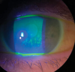
This group affects the epithelium and the tissue just beneath it, and Meesmann dystrophy, linked to KRT3 and KRT12, shows tiny intraepithelial microcysts (see Meesmann corneal dystrophy). Lisch dystrophy produces whorled or band-shaped gray opacities at a similar level, while gelatinous drop-like dystrophy, caused by TACSTD2 mutations, forms subepithelial amyloid nodules.
TGFBI Dystrophies in the IC3D Classification
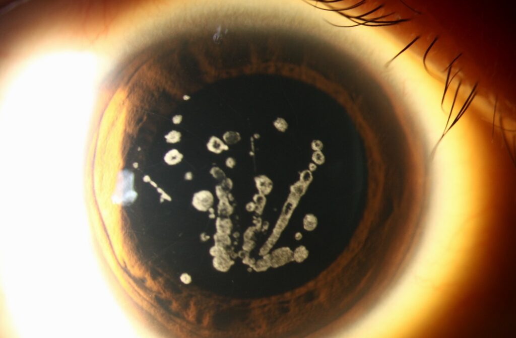
Mutations in TGFBI produce abnormal keratoepithelin deposits, and this group includes Reis-Bücklers, Thiel-Behnke, lattice type 1, granular type 1 and granular type 2. Clinicians formerly called granular type 2 Avellino dystrophy, and specific mutations often predict the phenotype; for example, R124H links to granular type 2. Therefore, genetic testing can clarify a confusing slit-lamp picture (see granular corneal dystrophy).
Stromal Dystrophies
These conditions primarily involve the stroma and spare the epithelium, and macular dystrophy follows autosomal recessive inheritance and involves CHST6. Schnyder dystrophy involves UBIAD1 and causes crystalline or lipid deposits, while congenital stromal, fleck, posterior amorphous, central cloudy of François and pre-Descemet dystrophies complete the group.
Endothelial Dystrophies
This group includes Fuchs, posterior polymorphous, congenital hereditary and X-linked endothelial dystrophies, and Fuchs dystrophy is by far the most common in clinic. Its genetics are heterogeneous, and a CTG repeat expansion in TCF4 accounts for many cases (see Fuchs endothelial corneal dystrophy). CHED involves SLC4A11 and follows recessive inheritance, and the second edition also retired the old dominant CHED1 label and folded it into posterior polymorphous dystrophy.
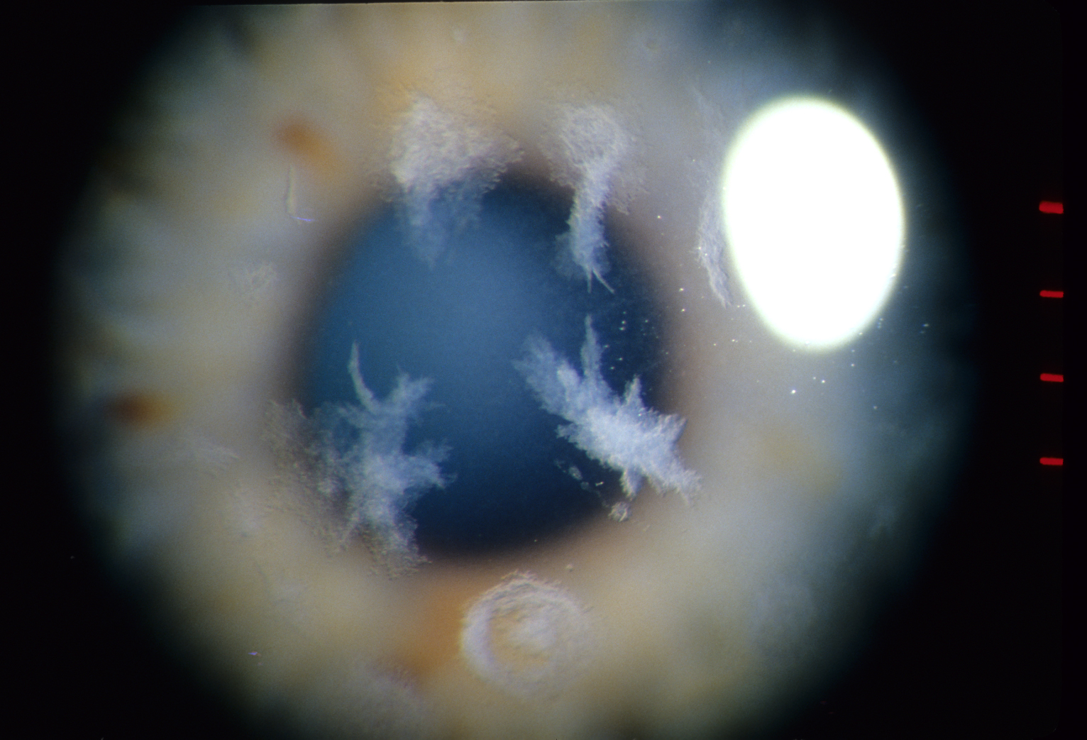
Evidence Categories in the IC3D Classification
The committee also grades how well each dystrophy is defined, and category 1 means a well-defined dystrophy with a known gene and specific mutations. In category 2, the dystrophy is well defined and mapped to a locus, while the gene remains unknown.
Category 3 covers a well-defined dystrophy that researchers have not yet mapped, while earlier editions also used category 4 for suspected dystrophies with weak evidence. Categories change as evidence grows, so quote them from the most recent edition.
Using the IC3D Classification at the Slit Lamp
Start with the layer and note whether opacities sit in the epithelium, at Bowman level, in the stroma or at the endothelium, then record pattern, clear intervals, laterality and age at onset. Next, ask about family history and recurrent erosions, and finally compare the findings with the template sheet for the most likely group.
Genetic Testing in Practice
Testing helps most with TGFBI dystrophies, where a single mutation often predicts the phenotype, and it also matters before refractive surgery because LASIK can worsen granular type 2 deposits. Results also support family counseling; however, a negative panel does not exclude a dystrophy, particularly in heterogeneous diseases such as Fuchs.
Limits of the IC3D Classification
The system describes current knowledge, which remains incomplete, and some phenotypes overlap, so one patient may not fit any template neatly. The anatomic grouping also simplifies diseases that cross layers; therefore, use it as a shared language, and keep clinical judgment in charge.
Discussing IC3D Classification With Patients
Patients benefit from knowing the layer involved, the inheritance and the expected course. For example, explain that many stromal dystrophies can recur after keratoplasty, and then explain which relatives should have a slit-lamp exam.
Pitfalls to Avoid
- Do not use outdated eponyms such as “Avellino” or “CHED1” in new reports
- Do not label every map-dot-fingerprint pattern as an inherited dystrophy
- Avoid LASIK in suspected TGFBI dystrophy before genetic testing
- Do not assume a negative gene panel excludes a dystrophy
- Remember that categories change between editions


Document what you see
Two smartphone imaging tools built for everyday clinic use — one for the slit lamp, one for the fundus.
From Choroida — the team behind this siteReferences
- Weiss JS, Møller HU, Lisch W, et al. The IC3D classification of the corneal dystrophies. Cornea. 2008;27(Suppl 2):S1-S83.
- Weiss JS, Møller HU, Aldave AJ, et al. IC3D classification of corneal dystrophies, edition 2. Cornea. 2015;34:117-159.
- Weiss JS, Rapuano CJ, Seitz B, et al. IC3D classification of corneal dystrophies, edition 3. Cornea. 2024;43:466-527.
- Wieben ED, Aleff RA, Tosakulwong N, et al. A common trinucleotide repeat expansion within the transcription factor 4 (TCF4, E2-2) gene predicts Fuchs corneal dystrophy. PLoS One. 2012;7:e49083.
Test yourself
A few questions straight from this article.
-
Under the IC3D classification, the old label Reis-Bücklers was split into Reis-Bücklers and which other Bowman layer dystrophy?
Appearance-based names had lumped two separate Bowman layer dystrophies under Reis-Bücklers; the IC3D committee separated them into Reis-Bücklers and Thiel-Behnke dystrophies. -
What did the 2015 second edition of the IC3D classification create in place of the Bowman layer group?
Because one gene, TGFBI, drives several dystrophies that span layers, the second edition replaced the Bowman layer group with an epithelial-stromal TGFBI group. -
Which genes does the IC3D classification link to Meesmann corneal dystrophy with its intraepithelial microcysts?
Meesmann dystrophy sits in the epithelial and subepithelial group, is linked to KRT3 and KRT12, and shows tiny intraepithelial microcysts. -
Which TGFBI mutation is linked to granular corneal dystrophy type 2, formerly called Avellino dystrophy?
Within the TGFBI group, specific mutations often predict the phenotype; R124H links to granular type 2, the dystrophy once called Avellino. -
How does the IC3D classification describe the inheritance and gene of macular corneal dystrophy?
Macular dystrophy is a stromal dystrophy that follows autosomal recessive inheritance and involves the CHST6 gene. -
Which genetic change accounts for many cases of Fuchs endothelial corneal dystrophy in the IC3D classification?
Fuchs genetics are heterogeneous, but a CTG repeat expansion in TCF4 accounts for many cases. -
What happened to the old dominant CHED1 label in the second edition of the IC3D classification?
The second edition retired CHED1 and folded it into posterior polymorphous dystrophy; CHED itself now refers to the recessive SLC4A11 disease. -
In the IC3D evidence grading, what defines a category 2 corneal dystrophy?
Category 1 has a known gene and mutations, category 2 is mapped to a locus with the gene still unknown, and category 3 has not yet been mapped. -
Why does genetic testing matter before refractive surgery in a suspected TGFBI corneal dystrophy?
Testing helps most in TGFBI disease, and LASIK should be avoided before testing because it can worsen granular type 2 deposits. -
When applying the IC3D classification at the slit lamp, what should the examiner establish first?
Start with the layer — epithelium, Bowman level, stroma or endothelium — then record pattern, clear intervals, laterality and onset before comparing with the template sheet.

Writes and reviews the clinical articles on Educate. Every post is checked against peer-reviewed sources before it goes live.