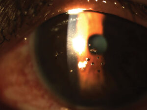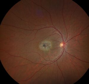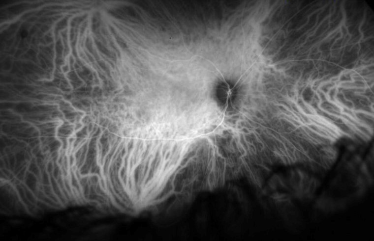
Pachyvessels are dilated large choroidal vessels, usually veins, located in Haller layer.
OCT shows them as large hyporeflective lumens beneath a thinned inner choroid. However, no universal size cutoff defines the sign, so interpretation requires judgment.
What Pachyvessels Look Like on OCT
Each vessel appears as a round or oval dark lumen in the outer choroid, and the surrounding stroma looks brighter, which helps outline the vessel. Above it, Sattler layer and the choriocapillaris often appear thinned. As a result, the large lumen seems to sit close to Bruch membrane.
Why Haller Layer Vessels Dilate
The exact mechanism remains uncertain, but one hypothesis proposes venous congestion within the vortex drainage system. For example, researchers have described macular vortex vein remodeling in pachychoroid eyes. Peripheral venous dilation has a separate clinical form (see vortex vein varix).
Fundus Explorer Pro
Photograph the retinal findings described here with the phone already in your pocket — 22 D optics and built-in illumination in one handheld unit.
From Choroida — the team behind this siteChoosing the Right Scan in Pachyvessels
Standard spectral domain scans may lose signal before reaching the outer choroid, so use enhanced depth imaging or swept-source OCT whenever possible. Dense raster scans also help because a single line can miss a focal dilation.
Reading a B-Scan for Pachyvessels
First, find the choroidoscleral junction and confirm that the outer choroid is visible, and next, compare the suspected vessel with neighboring lumens at the same depth. Then, examine the inner choroid above it for thinning or compression. Finally, scroll through adjacent scans to confirm a continuous tubular structure.
No Agreed Size Threshold
Published descriptions vary, and no single caliber separates normal from pathologic vessels. Therefore, avoid labeling a vessel from one diameter measurement, because a descriptive report is often safer than a firm numerical claim.
Choroidal Thickness and Pachyvessels
Choroidal thickness and vessel caliber are related, although they are not interchangeable. For example, a dilated vessel can cause only focal thickening in an otherwise average choroid. Moreover, thickness varies with age, axial length and time of day, so a normal subfoveal measurement does not exclude the sign.
En Face Confirmation of Pachyvessels
En face imaging shows the vessel course across the posterior pole (see en face OCT). True dilated veins usually appear as continuous channels that follow a recognizable path. However, poor segmentation can create false patterns, so always check the slab position on the B-scan.
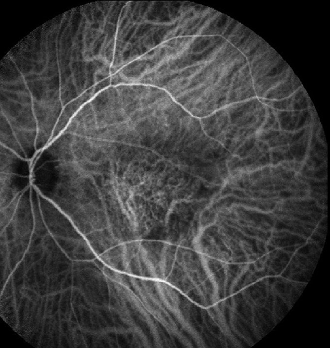
ICG and OCT Angiography
Indocyanine green angiography displays large choroidal veins directly, but it is invasive, so reserve it for questions that could change management. OCT angiography can assess the choriocapillaris above a dilated vessel (see OCT angiography). Because signal attenuation limits deep flow assessment, use structural OCT to identify the vessel.
Normal Variants That Mimic Pachyvessels
Healthy eyes can show one prominent Haller layer vessel beneath the macula, so a large lumen alone does not indicate disease. In addition, look for inner choroidal attenuation directly above the vessel, because its absence should lower your confidence in the sign.
Scan Geometry Errors
A vessel cut along its length can appear unusually long and wide, and similarly, an oblique section can exaggerate the apparent diameter of a round vessel. For these reasons, confirm the finding across several scans and on en face imaging.
Other Causes of a Thick or Dark Choroid
Inflammatory and infiltrative disease can thicken the choroid without a focal dilated vein. In addition, choroidal caverns can appear as dark spaces that resemble vessel lumens, so consider these alternatives when the appearance lacks a tubular course.
Reporting and Follow-Up of Pachyvessels
State the scan mode, vessel location, extent and any inner choroidal attenuation, then describe overlying retinal pigment epithelium changes or fluid (see subretinal fluid on OCT). An isolated dilated vessel in an asymptomatic eye does not itself call for treatment. When comparing visits, use the same device and scan protocol, and also advise prompt review for new blur, distortion or a central scotoma.
Pitfalls to Avoid
- Do not diagnose the sign when the outer choroid is poorly imaged
- Avoid relying on one B-scan or one diameter measurement
- Do not treat choroidal thickness as a substitute for vessel assessment
- Never accept an en face pattern without checking its segmentation
- Avoid assigning a disease label from the vessel alone


Document what you see
Two smartphone imaging tools built for everyday clinic use — one for the slit lamp, one for the fundus.
From Choroida — the team behind this siteReferences
- Cheung CMG, Lee WK, Koizumi H, Dansingani K, Lai TYY, Freund KB. Pachychoroid disease. Eye (Lond). 2019;33:14-33.
- Dansingani KK, Balaratnasingam C, Naysan J, Freund KB. En face imaging of pachychoroid spectrum disorders with swept-source optical coherence tomography. Retina. 2016;36:499-516.
- Spaide RF, Koizumi H, Pozzoni MC. Enhanced depth imaging spectral-domain optical coherence tomography. Am J Ophthalmol. 2008;146:496-500.
- Margolis R, Spaide RF. A pilot study of enhanced depth imaging optical coherence tomography of the choroid in normal eyes. Am J Ophthalmol. 2009;147:811-815.
- Matsumoto H, Kishi S, Mukai R, Akiyama H. Remodeling of macular vortex veins in pachychoroid neovasculopathy. Sci Rep. 2019;9:14689.
- Tan CS, Ouyang Y, Ruiz H, Sadda SR. Diurnal variation of choroidal thickness in normal, healthy subjects measured by spectral domain optical coherence tomography. Invest Ophthalmol Vis Sci. 2012;53:261-266.
Test yourself
A few questions straight from this article.
-
Pachyvessels are dilated large choroidal vessels, usually veins, located in which choroidal layer?
Pachyvessels are dilated large choroidal vessels, usually veins, in Haller layer. Above them, Sattler layer and the choriocapillaris often look thinned. -
How does a pachyvessel typically appear on an OCT B-scan?
Each vessel appears as a round or oval hyporeflective lumen in the outer choroid. The brighter surrounding stroma helps outline it, and the lumen seems to sit close to Bruch membrane. -
What vessel-diameter threshold defines a pachyvessel on OCT?
Published descriptions vary and no single caliber separates normal from pathological vessels. A descriptive report is often safer than labelling a vessel from one diameter. -
Which hypothesis has been proposed to explain the dilation of Haller layer vessels seen as pachyvessels?
The exact mechanism is uncertain, but one hypothesis proposes venous congestion in the vortex drainage system. Macular vortex vein remodelling has been described in pachychoroid eyes. -
Which OCT acquisition is preferred when assessing suspected pachyvessels?
Standard spectral-domain scans may lose signal before reaching the outer choroid, so enhanced depth imaging or swept-source OCT is preferred. Dense raster scans help because a single line can miss a focal dilation. -
Why does a normal subfoveal choroidal thickness not exclude pachyvessels?
Thickness and vessel caliber are related but not interchangeable; a dilated vessel can cause only focal thickening. Thickness also varies with age, axial length and time of day. -
Healthy eyes can show one prominent Haller vessel. Which OCT feature above the vessel supports a true pachyvessel?
A large lumen alone does not indicate disease. Look for inner choroidal attenuation directly above the vessel, because its absence should lower confidence in the sign. -
Choroidal caverns can resemble pachyvessel lumens on OCT. Which feature favours a true dilated vein?
Choroidal caverns can appear as dark spaces resembling lumens, so consider them when the appearance lacks a tubular course. Scroll through adjacent scans to confirm a continuous tubular structure. -
Why is structural OCT, rather than OCT angiography, used to identify a pachyvessel?
OCT angiography can assess the choriocapillaris above a dilated vessel, but signal attenuation limits deep flow assessment. Structural OCT is therefore used to identify the vessel itself. -
How should an isolated pachyvessel in an asymptomatic eye be managed?
An isolated dilated vessel in an asymptomatic eye does not itself call for treatment. Compare visits with the same device and protocol, and advise prompt review for new blur, distortion or a central scotoma.

Writes and reviews the clinical articles on Educate. Every post is checked against peer-reviewed sources before it goes live.