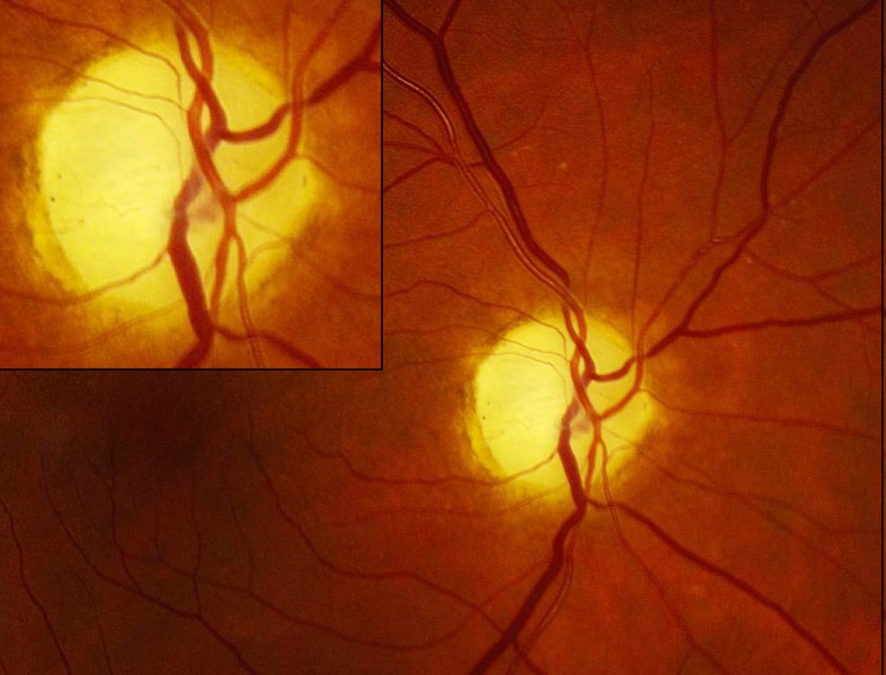
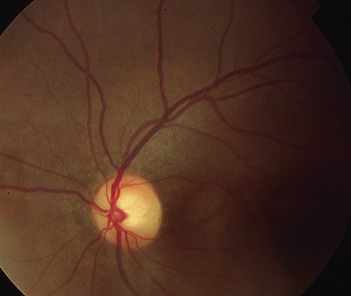
Bow-tie atrophy is a horizontal band of pallor across the optic disc, with relative sparing of the superior and inferior poles.
Clinicians also call it band optic atrophy, and the sign reflects loss of crossing nasal retinal fibers. Therefore, it points toward the chiasm or optic tract and usually calls for neuroimaging.
What the Examiner Sees in Bow-Tie Atrophy
Pallor crosses the nasal and temporal disc, while the upper and lower rims stay relatively pink, and the arcuate bundles often remain visible above and below the disc. However, early cases look subtle because the temporal disc normally appears paler.
The Fiber Anatomy Behind the Band
Nasal retinal ganglion cells send axons that cross at the chiasm. First, nasal macular and peripheral fibers enter the temporal and nasal sides of the disc, while temporal fibers, by contrast, arch around the macula and enter mainly at the poles. As a result, loss of crossed fibers removes the nasal and temporal disc tissue.
Fundus Explorer Pro
Photograph the retinal findings described here with the phone already in your pocket — 22 D optics and built-in illumination in one handheld unit.
From Choroida — the team behind this siteWhy Bow-Tie Atrophy Appears Late
Retrograde degeneration needs weeks to reach the disc, so a recent compressive lesion can produce a clear field defect with a normal disc. For this reason, a healthy-looking nerve never excludes chiasmal disease.
What the Patient Reports
Many patients notice little until central acuity falls or the fields narrow considerably, while others describe bumping into objects or trouble reading. Bitemporal loss can also cause hemifield slide with intermittent diplopia. In addition, ask about headache, menstrual changes, and galactorrhea.
Bow-Tie Atrophy in Chiasmal Lesions
Central chiasmal damage interrupts crossing fibers from both eyes, so both discs can show the band pattern, with bitemporal hemianopia on perimetry. Asymmetry is common because tumors rarely compress the chiasm evenly.
Unilateral Bow-Tie Atrophy in Optic Tract Lesions
A tract lesion damages crossed fibers from the contralateral eye and uncrossed fibers from the ipsilateral eye, and therefore the contralateral eye develops the band pattern. The ipsilateral eye loses temporal fibers, so its poles thin.
Fields show a homonymous hemianopia, which is often incongruous, and a relative afferent pupillary defect also tends to appear in the contralateral eye.
Congenital Retrogeniculate Lesions
Long-standing congenital lesions behind the geniculate can produce a similar disc pattern through trans-synaptic degeneration, and Hoyt and Kommerell described these fundus changes in homonymous hemianopia. By contrast, occipital lesions acquired in adulthood rarely cause obvious pallor.
OCT in Bow-Tie Atrophy
Peripapillary OCT often shows nasal and temporal thinning with preserved superior and inferior sectors, but average thickness may hide this pattern, so read the clock-hour maps.
Monteiro and colleagues found that OCT could detect band atrophy, although its classification had limits. In addition, macular ganglion cell analysis may show binasal thinning in chiasmal compression.
Differential Diagnosis of Disc Pallor
Glaucoma usually affects the poles first and enlarges the cup, while isolated temporal pallor suggests dominant optic atrophy.
Similarly, toxic and nutritional optic neuropathy produces central or cecocentral scotomas. Other causes of a pale disc also need consideration (see optic atrophy).
Causes of Bow-Tie Atrophy
Pituitary adenoma is a common cause in adults, while craniopharyngioma and meningioma also compress the chiasm and aneurysm is a dangerous alternative. Tract lesions may follow tumor, demyelination, stroke, or trauma.
Workup After the Sign Appears
First, request contrast-enhanced MRI that includes the sellar region, and tell the radiologist the suspected site, because routine sequences can miss small sellar lesions.
Then, add automated perimetry and check whether defects respect the vertical meridian in each eye. Order a pituitary hormone evaluation when a sellar process is suspected.
Management and Prognosis in Bow-Tie Atrophy
Visible atrophy signals established axonal loss, so recovery may be incomplete. However, Danesh-Meyer and colleagues found that preoperative nerve fiber layer thickness predicted visual recovery after parachiasmal tumor surgery.
No treatment reverses the atrophy, so management targets the causative lesion. Options include transsphenoidal surgery, dopamine agonists for prolactinoma, and radiotherapy.
Neurosurgery and endocrinology should share the plan. Explain that treatment aims to stop further damage, while lost fibers will not regrow.
Follow-Up and Counseling
Serial fields and OCT track recovery or recurrence, and new field loss after treatment should prompt repeat imaging. Also discuss driving, because bitemporal defects may affect legal fitness.
Pitfalls to Avoid
- Do not label band pallor as glaucoma without checking the vertical meridian
- Never let a normal disc end the workup for a bitemporal defect
- Do not rely on average OCT thickness alone
- Compare both discs in suspected tract lesions


Document what you see
Two smartphone imaging tools built for everyday clinic use — one for the slit lamp, one for the fundus.
From Choroida — the team behind this siteReferences
- Unsöld R, Hoyt WF. Band atrophy of the optic nerve: the histology of temporal hemianopsia. Arch Ophthalmol. 1980;98:1637-1638.
- Hoyt WF, Kommerell G. Der Fundus oculi bei homonymer Hemianopie. Klin Monbl Augenheilkd. 1973;162:456-464.
- Monteiro ML, Moura FC, Medeiros FA. Diagnostic ability of optical coherence tomography with a normative database to detect band atrophy of the optic nerve. Am J Ophthalmol. 2007;143:896-899.
- Danesh-Meyer HV, Papchenko T, Savino PJ, et al. In vivo retinal nerve fiber layer thickness measured by optical coherence tomography predicts visual recovery after surgery for parachiasmal tumors. Invest Ophthalmol Vis Sci. 2008;49:1879-1885.
- Tieger MG, Hedges TR, Ho J, et al. Ganglion cell complex loss in chiasmal compression by brain tumors. J Neuroophthalmol. 2017;37:7-12.
Test yourself
A few questions straight from this article.
-
What disc appearance defines bow-tie (band) optic atrophy?
Bow-tie atrophy is a horizontal band of pallor across the nasal and temporal disc, while the upper and lower rims stay relatively pink. -
Loss of which retinal ganglion cell fibers produces bow-tie optic atrophy?
The sign reflects loss of the crossing nasal retinal fibers, which is why it points toward the chiasm or optic tract and usually calls for neuroimaging. -
Why can a recent chiasmal compression produce a clear bitemporal field defect while the disc still looks normal?
Retrograde degeneration takes weeks to reach the disc, so a healthy-looking nerve never excludes chiasmal disease. -
Which perimetric finding accompanies bilateral bow-tie atrophy from central chiasmal damage?
Central chiasmal damage interrupts crossing fibers from both eyes, so both discs can show the band and perimetry shows bitemporal hemianopia, often asymmetric because tumors rarely compress evenly. -
In an optic tract lesion, which eye develops the bow-tie atrophy pattern?
A tract lesion damages crossed fibers from the contralateral eye, which develops the band pattern, while the ipsilateral eye loses temporal fibers so its poles thin. -
Which visual symptom can bitemporal field loss from chiasmal lesions with bow-tie atrophy cause?
Bitemporal loss can cause hemifield slide with intermittent diplopia; others notice little until acuity falls, or report bumping into objects or trouble reading. -
Which retrogeniculate lesion can produce a bow-tie disc pattern through trans-synaptic degeneration?
Long-standing congenital retrogeniculate lesions can cause a similar disc pattern via trans-synaptic degeneration, whereas occipital lesions acquired in adulthood rarely cause obvious pallor. -
When reading peripapillary OCT in suspected bow-tie atrophy, why should clock-hour maps be checked?
OCT often shows nasal and temporal thinning with preserved superior and inferior sectors, but average thickness can mask this, so the clock-hour maps should be read. -
What should the first imaging request include once bow-tie atrophy is found?
Request contrast-enhanced MRI that covers the sellar region and tell the radiologist the suspected site, because routine sequences can miss small sellar lesions. -
Which measurement predicted visual recovery after surgery for parachiasmal tumors in patients at risk of bow-tie atrophy?
Visible atrophy means established axonal loss and incomplete recovery is possible, but preoperative nerve fiber layer thickness predicted visual recovery after parachiasmal tumor surgery.

Writes and reviews the clinical articles on Educate. Every post is checked against peer-reviewed sources before it goes live.