Nonglaucomatous cupping is optic disc excavation caused by a disease other than glaucoma.
Compressive lesions, ischemia and hereditary neuropathies can all produce a large cup. However, the rim, the visual field and the history usually separate these conditions from glaucoma.
Why the Cup Enlarges Without Glaucoma
Any cause of axon loss can thin the rim and enlarge the cup. Glaucoma adds backward bowing of the lamina cribrosa, which deepens the excavation. In contrast, many other optic neuropathies cause pallor with less laminar change.
Rim Color in Nonglaucomatous Cupping
Rim color is the most useful single sign at the slit lamp. In glaucoma, the remaining rim usually stays pink, even when it is thin, whereas pallor of the remaining rim points toward a nonglaucomatous process.
Trobe and colleagues reported that rim pallor favored nonglaucomatous disease, while focal rim loss favored glaucoma. Compare both eyes as well, because asymmetric pallor stands out against a healthier fellow disc.
Fundus Explorer Pro
Photograph the retinal findings described here with the phone already in your pocket — 22 D optics and built-in illumination in one handheld unit.
From Choroida — the team behind this siteRim Shape and Cup Depth
Glaucoma typically erodes the superior and inferior poles, which elongates the cup vertically. Notches, disc hemorrhages and nerve fiber bundle defects then support that diagnosis.
By contrast, a shallow cup with a pale, relatively preserved rim should raise suspicion. However, no single disc sign is definitive, so correlate the appearance with function.
Visual Field Patterns in Nonglaucomatous Cupping
Glaucoma usually produces arcuate defects, nasal steps and paracentral scotomas that respect the horizontal midline. In contrast, defects that respect the vertical midline suggest a chiasmal or retrochiasmal lesion.
Central or cecocentral scotomas also suggest another diagnosis, such as a hereditary or toxic neuropathy. Therefore, inspect the vertical meridian in both eyes on every field.
Acuity and Color Vision in Nonglaucomatous Cupping
Glaucoma usually spares central acuity until late in the disease, so reduced acuity with a modest cup needs an explanation. Greenfield and colleagues linked younger age, lower acuity, vertically aligned defects and pallor exceeding cupping with nonglaucomatous disease, so test color vision and check for a relative afferent pupillary defect.
Compressive Causes of Nonglaucomatous Cupping
Pituitary adenomas, meningiomas, craniopharyngiomas and carotid aneurysms can compress the anterior visual pathway. Slow compression may cause gradual, painless field loss that resembles normal-tension glaucoma, and the disc may show pallor, true cupping or both.
As a result, review atypical low-pressure cases carefully before starting drops. Interpret the pressure alongside corneal pachymetry, because corneal thickness alters the reading.
Ischemic Optic Neuropathy and the Late Cup
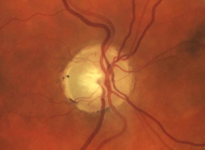
Arteritic anterior ischemic optic neuropathy can leave a large cup with marked pallor. Hayreh and Jonas described this cupping after arteritic disease, while nonarteritic eyes usually lacked comparable excavation. Therefore, ask about prior sudden visual loss, jaw claudication and scalp tenderness.
Hereditary, Toxic and Traumatic Causes
Leber hereditary optic neuropathy and dominant optic atrophy can leave temporal pallor, sometimes with cupping. Methanol toxicity and optic nerve trauma can also produce excavation. In addition, chronic papilledema can leave a pale atrophic disc (see idiopathic intracranial hypertension).
Large Discs and Congenital Mimics
Large optic discs naturally have large cups, so estimate disc size before judging the ratio. A physiologic big cup keeps a healthy pink rim and stays stable over time. Optic pits, colobomas and tilted discs can also imitate excavation.
OCT in Nonglaucomatous Cupping
OCT quantifies thinning, although it cannot identify the cause alone. Binasal ganglion cell loss that respects the vertical midline suggests chiasmal disease, while glaucoma tends to affect the superior and inferior poles first. However, advanced damage from any cause can flatten these patterns.
When Nonglaucomatous Cupping Needs Imaging
Consider contrast-enhanced MRI of the brain and orbits when these features appear:
- Rim pallor exceeding the degree of cupping
- Field defects that respect the vertical midline
- Acuity worse than the disc explains, especially below 20/40
- Younger age, particularly under 50, with unexplained cupping
- Progression despite well-controlled pressure
Treatment of Nonglaucomatous Cupping
Treatment targets the cause, so an accurate diagnosis directs every later decision. For example, decompression can improve vision in compressive disease, although recovery varies.
Corticosteroids aim to protect the fellow eye in giant cell arteritis, yet they rarely restore lost vision. In contrast, pressure-lowering drops have no established benefit without glaucoma, and they add cost and side effects.
Counseling Patients About Nonglaucomatous Cupping
Explain that a large cup describes the nerve’s appearance, and several conditions can cause it. Also explain that imaging looks for treatable causes, so a normal scan still helps. Then warn patients that existing atrophy may remain, even after the cause resolves.
Pitfalls to Avoid
- Do not diagnose normal-tension glaucoma before reviewing rim color and the vertical meridian
- Never judge the disc by cup-to-disc ratio alone
- Do not dismiss a bitemporal field as artifact without repeating it


Document what you see
Two smartphone imaging tools built for everyday clinic use — one for the slit lamp, one for the fundus.
From Choroida — the team behind this siteReferences
- Greenfield DS, Siatkowski RM, Glaser JS, Schatz NJ, Parrish RK. The cupped disc: who needs neuroimaging? Ophthalmology. 1998;105:1866-1874.
- Trobe JD, Glaser JS, Cassady J, Herschler J, Anderson DR. Nonglaucomatous excavation of the optic disc. Arch Ophthalmol. 1980;98:1046-1050.
- Hayreh SS, Jonas JB. Optic disc morphology after arteritic anterior ischemic optic neuropathy. Ophthalmology. 2001;108:1586-1594.
- Fraser CL, White AJ, Plant GT, Martin KR. Optic nerve cupping and the neuro-ophthalmologist. J Neuroophthalmol. 2013;33:377-389.
Test yourself
A few questions straight from this article.
-
Which structural change does glaucoma add to axon loss that deepens the excavation compared with nonglaucomatous cupping?
Any axon loss thins the rim and enlarges the cup, but glaucoma adds backward bowing of the lamina cribrosa, whereas many other optic neuropathies cause pallor with less laminar change. -
Which single slit-lamp sign is most useful for separating nonglaucomatous cupping from glaucoma?
In glaucoma the remaining rim usually stays pink even when thin, whereas pallor of the remaining rim points toward a nonglaucomatous process. -
Which rim pattern favors glaucoma over nonglaucomatous cupping?
Glaucoma typically erodes the superior and inferior poles, elongating the cup vertically, with notches, disc hemorrhages and nerve fiber bundle defects supporting the diagnosis. -
In a patient with optic disc cupping, which visual field feature suggests a chiasmal or retrochiasmal lesion rather than glaucoma?
Glaucoma produces arcuate defects, nasal steps and paracentral scotomas respecting the horizontal midline; defects respecting the vertical midline suggest a chiasmal or retrochiasmal lesion. -
In unexplained optic disc cupping, below which age is the finding a red flag that supports contrast-enhanced MRI?
MRI of the brain and orbits is advised for younger patients, particularly under 50, with unexplained cupping, alongside acuity below 20/40, vertical-midline defects and rim pallor exceeding cupping. -
Slow compression of the anterior visual pathway can produce cupping and gradual painless field loss that mimics which condition?
Pituitary adenomas, meningiomas, craniopharyngiomas and carotid aneurysms can cause gradual painless field loss resembling normal-tension glaucoma, so atypical low-pressure cases need careful review before drops. -
Which form of ischemic optic neuropathy can leave a large cup with marked pallor that mimics glaucomatous cupping?
Arteritic disease can leave a large, markedly pale cup, while nonarteritic eyes usually lack comparable excavation, so ask about prior sudden loss, jaw claudication and scalp tenderness. -
Which finding supports a physiologic big cup rather than nonglaucomatous cupping?
Large discs naturally have large cups, so disc size should be estimated first; a physiologic big cup keeps a healthy pink rim and remains stable over time. -
In giant cell arteritis causing nonglaucomatous cupping, what is the main aim of corticosteroid treatment?
Corticosteroids aim to protect the fellow eye in giant cell arteritis, yet they rarely restore vision already lost. -
What is the role of pressure-lowering drops in nonglaucomatous cupping?
Treatment targets the cause; pressure-lowering drops have no established benefit without glaucoma and only add cost and side effects.

Writes and reviews the clinical articles on Educate. Every post is checked against peer-reviewed sources before it goes live.